Shear Forces at the Heel: The Hidden Damage Mechanism
When clinicians think about pressure ulcer prevention, they typically focus on pressure—the perpendicular force that compresses tissue against a surface. This is understandable. The condition is called a pressure ulcer. Pressure is in the name.
But pressure tells only part of the story. Shear—the force acting parallel to the support surface that distorts tissue layers relative to each other—may be even more damaging than pressure alone. Research dating back to Bennett's seminal 1979 study demonstrated that when shear is present, the pressure required to produce vascular occlusion is reduced by approximately 50%. At shear levels of roughly 100 g/cm², the pressure needed to stop blood flow was half that required when little shear was present.
This finding has profound implications for heel protection. A device that reduces pressure but doesn't address shear may leave the heel vulnerable to the very damage it was meant to prevent.
What shear actually is.
It's easiest to imagine shear force acting parallel to a support surface, not perpendicular to it.
Imagine placing your hand flat on a table and pressing down—that's pressure. Now imagine sliding your hand along the table surface while pressing down—the friction between your skin and the table is resisting shear force.
At the tissue level, shear doesn't require gross movement. When the heel rests on a surface:
- The skin adheres to the surface through friction
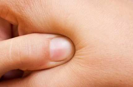
Whenever tissue is deformed like this both pressure and shear are present and the effects are not just surface effects
- The skeleton (calcaneus) may shift slightly relative to the skin
- The soft tissue between skin and bone is stretched and distorted
- Blood vessels running through this tissue are stretched and compressed
This *internal* distortion is shear; it's not just a surface effect. It happens even when the patient appears stationary. As Reichel first identified in 1958, shear causes subepidermal blood vessels to bend at right angles—a finding that remains fundamental to our understanding of shear-related tissue damage.
Shear damages tissue through vascular occlusion.
The mechanism by which shear causes harm is vascular—it compromises blood flow.
Blood vessels in soft tissue don't run perfectly perpendicular to the skin surface. Many run at angles, threading between tissue layers. When those layers shift relative to each other:
- Vessels are stretched longitudinally
- Vessel walls are compressed as they're pulled taut
- Blood flow through the stretched, compressed vessels is reduced or stopped
- Tissue downstream of the occlusion becomes ischaemic (oxygen-deprived)
Bennett's research demonstrated that shear forces are extremely effective at stopping blood flow—more so than perpendicular pressure alone. Shear causes what he described as "stretching and tearing of tiny blood vessels," reducing blood flow through a fundamentally different mechanism than simple compression.
One way to understand this: imagine a garden hose lying on the ground. You could stop water flow by stepping directly on the hose (pressure), or by pulling the hose taut while it's pinched under something (shear) or by putting a kink into the hose. The second method can be equally effective with less direct force.
Shear achieves vascular occlusion with less obvious external force than perpendicular pressure, making it a hidden contributor to tissue damage.
The heel is particularly susceptible to shear.

The heel's anatomy creates conditions where shear is especially problematic. This isn't speculation—the heel is the second most common site for pressure ulcer development after the sacrum, according to systematic reviews of prevalence data. In critical care settings, 12.8% of patients develop deep-tissue pressure injuries, most commonly at the heel.
Thin tissue layers. There's minimal soft tissue between the skin and the calcaneus. The tissue present is relatively avascular fat, easily distorted by applied forces.
Curved bony surface. The calcaneus isn't flat—it's a curved prominence. When the heel rests on a surface, the bone naturally wants to shift position, creating shear between the skin (adhered to the surface) and the bone (moving relative to it).
Blood supply. The blood supply to the heel originates from the posterior tibial and peroneal arteries, forming an arterial plexus at the terminal ends of their pathways. These are essentially end-arteries without redundant collateral supply. When shear occludes these vessels, there's no alternative blood flow pathway. Limited collateral circulation means reduced compensatory blood flow when vessels are compressed.
Dependent position in bed. The heel's typical position creates conditions where shear forces naturally develop—gravity pulls the leg downward, friction holds the skin in place, and the tissue between is caught in the middle.
Age-related changes. Tissue thinning with age further reduces the already minimal protection. In patients with peripheral arterial disease, vessels become stiff and cannot dilate effectively after pressure relief, compounding the vulnerability.
Shear occurs in ways that aren't always obvious.
Several common scenarios generate shear at the heel without necessarily being recognised as shear risks:
When a patient gradually slides toward the foot of the bed (common when the head of the bed is elevated), the skin may adhere to the sheet while the body moves. Shear forces develop throughout areas in contact with the bed, including the heels, unless a “slide sheet” is used.
When the head of the bed is raised or lowered, the mattress surface moves relative to the patient. Unless the patient is fully lifted clear of the surface during adjustment, shear forces act at the contact points.
Of course, if a patient is pulled across a surface rather than lifted, shear forces act on all tissue in contact with that surface.
Micromovement takes place during rest. Even a sedated patient isn't perfectly still. Small movements—breathing, involuntary shifts—can generate repeated low-level shear at the heel-surface interface.
Some pressure-redistributing mattresses have surfaces that move or shift to redistribute pressure. This movement can generate shear forces at the tissue interface.
Prevention strategies that focus only on perpendicular pressure may miss these shear-generating scenarios entirely.
Pressure and shear interact.
Shear doesn't act in isolation—it combines with pressure to create conditions worse than either alone.
When tissue is compressed (pressure) AND distorted (shear) simultaneously:
- Compression reduces the tissue's tolerance to distortion
- Distortion reduces the tissue's tolerance to compression
- The threshold for damage is lower than for either force in isolation
Research by Mak, Zhang and Tam in their 2010 Annual Review of Biomedical Engineering article identified that normal compression and shear deformation are the most important deformation modes in pressure ulcer formation. Their work showed that superficial pressure ulcers correlate with superficial shear forces, while deep tissue injuries correlate with perpendicular pressure combined with high deep shear near bony prominences.
The interaction is described in the literature as "particularly effective in promoting blood flow occlusion." The critical point is this: moderate pressure combined with moderate shear can produce tissue damage that neither would cause alone.
The heel, resting on a surface, typically experiences both forces simultaneously. Addressing one while ignoring the other leaves the tissue vulnerable.
Surface measurement doesn't capture shear damage.
One complication with shear is that its effects occur in deep tissue, often before surface changes are visible.
The tissue distortion from shear damages blood vessels in the subcutaneous layers—beneath the skin, not at its surface. Ischaemic damage begins in these deeper layers and may not become visible at the skin surface until substantial harm has occurred.
The research on deep tissue injury explains this phenomenon. Gefen and colleagues demonstrated that muscle tissue is substantially more susceptible to ischaemic damage than skin. High tissue deformations cause first signs of cell damage within minutes, but ischaemic damage takes several hours to fully develop. Oomens and colleagues, in their 2015 Annals of Biomedical Engineering paper "Pressure induced deep tissue injury explained," identified two sequential damage mechanisms: direct deformation (occurring in minutes) and ischaemic damage (developing over hours).
The first clinical sign—purple or maroon discolouration—appears approximately 48 hours after the pressure event. By the time surface ulceration appears, the wound has already involved the entire tissue depth.
This is why pressure ulcers sometimes appear "suddenly"—what looks like rapid surface breakdown may actually be the late manifestation of deep tissue damage that developed over days.
Pressure mapping devices, which measure interface pressure at the skin surface, don't capture shear forces or predict deep tissue damage from shear. A pressure map showing acceptable interface pressures may miss shear-related damage beneath the surface. Peak shear stresses, importantly, do not occur at the same location or time as peak pressures—which explains why pressure readings alone poorly predict ulceration sites.
Regular visual inspection of the heel is important, but the absence of surface changes doesn't guarantee tissue safety. Shear damage may be developing before it becomes visible.
Complete offloading eliminates both pressure and shear.
This is the critical insight for heel protection: true offloading—heel floating and not in contact with a support surface eliminates shear and pressure.
When the heel is suspended in space, contacting nothing:
- No perpendicular pressure acts on the tissue (nothing to compress against)
- No shear can develop (no surface for skin to adhere to)
- Both mechanisms of vascular compromise are prevented
This is fundamentally different from pressure reduction strategies that maintain heel contact with a softer surface. Foam, gel, and cushioning materials may reduce perpendicular pressure, but they still create an interface where shear can occur.
As long as the skin is in contact with any surface, shear remains possible. Only a complete suspension eliminates the problem.
What the research shows about device effectiveness.
The evidence base for heel protection devices continues to develop. A meta-analysis by Shi, Dumville, and Cullum, published in the International Wound Journal, found that foam and air mattresses show approximately a 50% reduction in relative risk compared to standard hospital mattresses. However, the systematic reviews note that the evidence for heel-specific devices remains limited, though the physiological rationale is strong.
Several studies provide encouraging data:
- An Australian randomised controlled trial of 394 patients found that heel suspension boots significantly reduced pressure injury compared to pillows. Notably, the intervention group experienced only superficial injuries, while the control group developed injuries up to stage 4.
- A comparison of suspension boots versus IV fluid bags (an improvised approach) showed 0% versus 40% pressure injury incidence (p=0.006).
- A trial of foam suspension boots in hip fracture surgery patients recorded 0 versus 29 heel pressure injuries (p<0.001).
The evidence supports what the biomechanics predicts: devices that achieve complete heel suspension outperform those that merely cushion.
There are practical considerations. Research indicates compliance challenges with some devices—32% of patients report sleep interference, and 41% report movement restriction. These factors matter in clinical practice and device selection.
Implications for device selection.
Understanding shear changes how we evaluate heel protection devices:
Foam boots and cushions may reduce interface pressure to some degree but maintain contact where shear can occur. They address one mechanism but not the other.
Gel-based products face the same limitation—the heel still contacts a surface, even a soft deformable one.
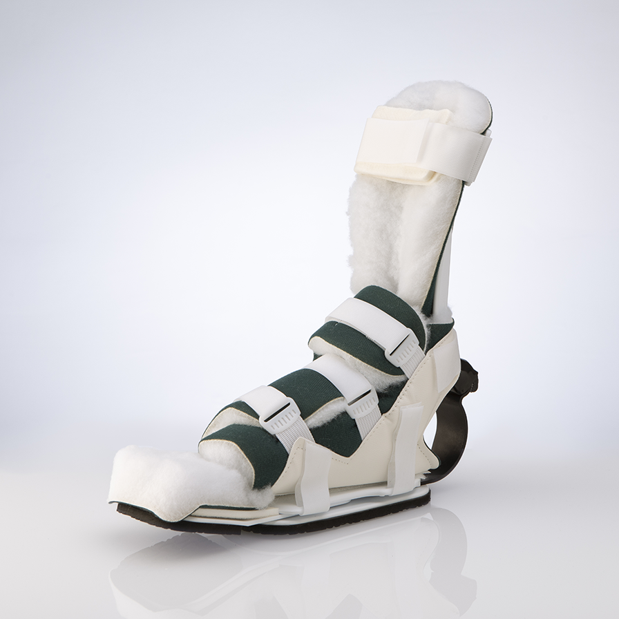
The 652SKG PRAFO design
Pillows allow shear whenever the heel contacts the pillow surface or shifts across it. The current international guidelines are explicit: for stage 3 or greater injuries, only specifically designed heel suspension devices should be used—not pillows or foam.
Suspension devices that float the heel entirely clear eliminate both pressure and shear. No contact means no interface where either force can act.
The PRAFO designs achieve this through their suspension approach—the heel floats in the device, supported by calf and foot contact but with the heel itself touching nothing. Both mechanical damage pathways are addressed.
What the guidelines recommend.
The 2024 NPIAP/EPUAP/PPPIA International Pressure Injury Guideline is explicit on heel protection:
- Heels must be completely elevated and free from contact with bed surfaces
- For stage 3 or greater injuries: only specifically designed heel suspension devices (not pillows or foam)
- The knee should be flexed approximately 5-10 degrees
- Pressure should be distributed evenly along the calf
- Excessive pressure on the Achilles tendon and popliteal vein should be avoided
This recommendation carries a B1 evidence strength with a strong recommendation rating.
The guideline also notes that prophylactic dressings can reduce pressure injury occurrence, but should be adjuncts to offloading—not replacements for it. The primary intervention remains complete heel suspension.
The overlooked mechanism.
Shear remains underappreciated in routine pressure ulcer prevention. The focus on pressure—reasonable given the condition's name—can obscure the role of parallel forces in tissue damage.
At the heel specifically, with its thin tissue coverage, curved bony prominence, end-artery blood supply, and vulnerable anatomy, shear may be as important as pressure in the development of ulceration. The research supports this: Bennett's finding that shear halves the pressure threshold for vascular occlusion is not a minor modifier—it fundamentally changes the damage equation.
Prevention strategies that achieve pressure reduction while maintaining surface contact address only half the problem. The heel that rests on a pressure-reducing surface is still subject to shear whenever relative movement occurs between skin and surface.
True heel protection requires eliminating both forces. That means heel floating—complete suspension with no surface contact.
A complete solution addresses both mechanisms.
The international guideline recommendation that heels be "fully free from contact with the support surface" implicitly addresses both pressure and shear. Full freedom from contact means no perpendicular force and no interface for parallel force. Both damage mechanisms are prevented.
Devices designed to this standard—including the Prafo range—protect against the hidden mechanism as well as the obvious one.
When evaluating any heel protection approach, the question isn't just "does it reduce pressure?" but "does it eliminate contact?" Only complete offloading addresses both dimensions of mechanical tissue damage.
Shear may be the hidden mechanism, but its effects are real. Effective prevention must account for it.
References
Bennett L, Kavner D, Lee BK, Trainor FA. Shear vs Pressure as Causative Factors in Skin Blood Flow Occlusion. Archives of Physical Medicine and Rehabilitation. 1979;60:309-314.
Coleman S, Nixon J, Keen J, et al. Our contemporary understanding of the aetiology of pressure ulcers/pressure injuries. International Wound Journal. 2022;19(3):692–704. doi:10.1111/iwj.13667.
Gefen A, van Nierop B, Bader DL, Oomens CW. Strain-time cell-death threshold for skeletal muscle in a tissue-engineered model system for deep tissue injury. *Journal of Biomechanics*. 2008;41(9):2003-2012.
Mak AFT, Zhang M, Tam EWC. Biomechanics of Pressure Ulcer in Body Tissues Interacting with External Forces during Locomotion. *Annual Review of Biomedical Engineering*. 2010;12:29-53. DOI: 10.1146/annurev-bioeng-070909-105223.
Barakat‑Johnson M, Mistiaen P, Lai M, Stephenson J, Buhr H, Campbell J, et al. Efficacy of a heel offloading boot in reducing heel pressure injuries in patients in Australian intensive care units: a single‑blinded randomised controlled trial. Intensive and Critical Care Nursing. 2022;71:103231.
European Pressure Ulcer Advisory Panel, National Pressure Injury Advisory Panel and Pan Pacific Pressure Injury Alliance. Prevention and Treatment of Pressure Ulcers/Injuries: Clinical Practice Guideline. The International Guideline. Emily Haesler (Ed.). EPUAP/NPIAP/PPPIA; 2019.
Oomens CW, Bader DL, Loerakker S, Baaijens F. Pressure induced deep tissue injury explained. *Annals of Biomedical Engineering*. 2015;43(2):297-305.
Reichel SM. Shearing force as a factor in decubitus ulcers in paraplegics. JAMA. 1958 Feb 15;166(7):762‑763. DOI: 10.1001/jama.1958.62990070004010a.
Nicosia G, Gatti M, Giannini G, et al. The effect of pressure-relieving surfaces on the prevention of heel ulcers in a variety of settings: a meta-analysis. International Wound Journal. 2007;4(2):111–118.
Shi C, Dumville JC, Cullum N. Support surfaces for pressure ulcer prevention: a network meta-analysis. PLoS One. 2018;13(2):e0192707.
Shi C, Dumville JC, Cullum N, Rhodes S, McInnes E. Foam surfaces for preventing pressure ulcers. Cochrane Database of Systematic Reviews. 2021;Issue 5:CD011655.
Shi C, Dumville JC, Cullum N, et al. Beds, overlays and mattresses for preventing and treating pressure ulcers: an overview of Cochrane Reviews and network meta-analysis. 2021.
Tubaishat A, Papanikolaou P, Anthony D, Habiballah L. Pressure ulcers prevalence in the acute care setting: a systematic review, 2000-2015. *Clinical Nursing Research*. 2018; 27(6):643-659.
Black J. Ten top tips: pressure ulcers on the heels. Wounds International. 2018. Available at: https://woundsinternational.com