Transcutaneous Vagus Nerve Stimulation: An Approved Medical Device with Emerging Rehabilitation Promise
A CE-Marked Device with Four Approved Indications—and Significant Potential Beyond Them
The vagus nerve has become something of a celebrity in rehabilitation circles. Search online, and you'll find claims ranging from the plausible to the frankly miraculous. As someone who has worked with electrical stimulation for over fifty years, I know that sometimes what appears new and original is based on a lot of work that has gone quietly unnoticed. So let me share in this article some of what the evidence actually shows about transcutaneous vagus nerve stimulation (tVNS) and what it might mean for people recovering from neurological conditions. If you're not familiar with the vagus nerve and what it's responsible for, I'll explain some of that further down.
This article focuses partly on a specific device: the tVNS® system, manufactured by tVNS Technologies GmbH in Germany. This is not a wellness gadget or an unregulated consumer product. The tVNS device is approved as a Class IIa medical device under the EU Medical Device Regulation (EU-MDR) with CE marking—currently the only non-invasive VNS device with this level of EU-MDR approval. It is registered for four specific clinical indications: epilepsy, depression, chronic migraines, and Prader-Willi syndrome. Anatomical Concepts (UK) is delighted to distribute and support the tVNS® system in the UK.
Why does this matter for rehabilitation? Because the same mechanisms that make vagus nerve stimulation effective for these approved conditions—neuroplasticity enhancement and anti-inflammatory action—are precisely the mechanisms that show promise for neurological rehabilitation. The ongoing research into stroke recovery, spinal cord injury, multiple sclerosis, and other conditions builds on a foundation of established science and regulatory-grade engineering.
Let me explain what we know, what we're learning, and where the boundaries currently sit.
The tVNS Device: Regulatory Status and Approved Indications
A Class IIa Medical Device
The tVNS® device has achieved CE marking as a Class IIa medical device under the EU Medical Device Regulation. This classification is significant—it indicates the device has met rigorous regulatory standards for safety and performance, including clinical evaluation. Class IIa devices carry a moderate level of risk and require conformity assessment by a Notified Body, which means independent verification that the device performs as claimed and safely.
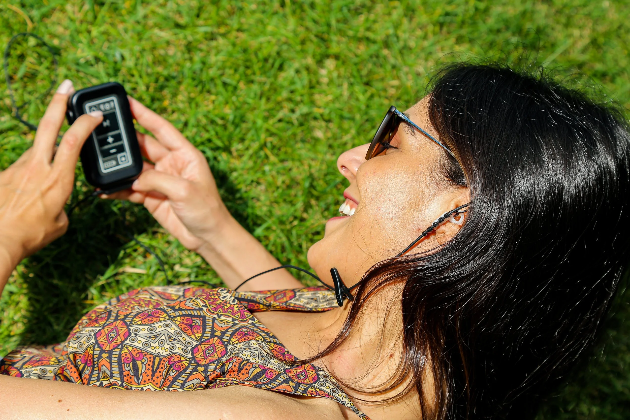
The device is manufactured in Germany and uses an electrode in the cymba concha of the ear—the inner, cup-shaped region—which is innervated 100% by the auricular branch of the vagus nerve (ABVN). This compares favourably with devices that stimulate the tragus, which achieve only approximately 45% vagal innervation. This anatomical precision matters: the cymba concha has the densest concentration of vagal afferent fibres in the external ear, meaning stimulation here provides the most direct pathway to the brainstem nuclei that drive the therapeutic effects.
tVNS system
The Four Approved Indications
The tVNS system is presently registered for four conditions, each supported by clinical evidence:
Epilepsy: Transcutaneous VNS has demonstrated clinical outcomes comparable to those of invasive (surgically implanted) VNS, without the surgical risks. Long-term data show a responder rate of approximately 30% (defined as patients achieving a 50% or greater reduction in seizure frequency), compared with 32% for implanted VNS. A 34.2% reduction in seizure frequency has been reported after 20 weeks of treatment. The comparable efficacy without surgical risk represents a meaningful advance for patients with drug-resistant epilepsy.
Depression: The vagus nerve's connections to brain regions involved in mood regulation—particularly through the locus coeruleus (norepinephrine) and raphe nuclei (serotonin)—provide the biological rationale for this indication. Clinical studies report 2–3 point reductions on the Hamilton Depression Rating Scale (HAMD) after 9 weeks of treatment. Implanted VNS has held FDA approval for treatment-resistant depression since 2005, and transcutaneous approaches now offer a non-invasive alternative.
Chronic Migraines: The trigemino-autonomic reflex, central to headache pathophysiology, can be modulated through vagal stimulation. Clinical evidence demonstrates a 50% reduction in migraine frequency after 12 weeks of tVNS treatment.
Prader-Willi Syndrome: Perhaps the most unexpected approved indication. This rare genetic condition involves chronic behavioural dysregulation, hyperphagia, and significant management challenges for families and carers. Clinical evidence shows 80% of patients achieved a 50% reduction in daily behavioural outbursts after 12 months of treatment—a substantial improvement in quality of life for both patients and their families.
KEY POINT: These four indications are covered under the device's medical device registration and reflect the current strength of the clinical evidence. The approved indications provide a regulatory and clinical foundation. What follows in this article—rehabilitation applications for stroke, spinal cord injury, MS, and other neurological conditions—represents emerging evidence that builds on the same underlying mechanisms but would currently be considered exploratory use of the device. Care must be taken to distinguish between approved and exploratory applications.
Why Vagus Nerve Stimulation Matters for Rehabilitation?
The Anatomy: A Wandering Nerve with Extraordinary Reach
The vagus nerve (cranial nerve X) extends from the brainstem down through the neck, chest, and abdomen, connecting the brain to the heart, lungs, gut, and numerous other organs. The name itself tells you something important—*nervus vagus* comes from the Latin for "wandering," and wander it does. No other cranial nerve has such an extensive reach.
The vagus nerve is the premier port of entry for interrogating the human nervous system. As the tenth cranial nerve, the vagus serves as a bidirectional superhighway, maintaining a constant dialogue between the brainstem and the visceral landscape. By delivering precise electrical pulses to this nerve non-invasively, we can "hack" the body's internal circuitry to treat disorders ranging from drug-resistant epilepsy to systemic inflammation.
Some years ago, I was involved in corporate development, teaching executives to manage stress by changing their breathing and their focus. This affected their heart rate variability and helped “balance” their autonomic nervous system. This meditation trick actually utilised the vagus nerve to achieve that balance in the autonomic nervous system. Fundamentally, the vagus nerve is a vital component of our health, and we are mostly oblivious to its presence.
A detail that matters for understanding tVNS:
Approximately 80% of vagal fibres are afferent—meaning they carry sensory information from the body organs to the brain.( Foley & DuBois, 1937) Only 20% are efferent (carrying signals outward). This 4:1 ratio explains why stimulating the vagus nerve can profoundly influence brain function. The vagus is not simply a "relaxation nerve" as popular accounts suggest. It is a bidirectional information highway between body and brain, with profound effects on neuroplasticity.
Two Complementary Mechanisms
How tVNS Reaches the Brain
The mechanism involves a cascade of events:
1. Electrical stimulation activates sensory nerve fibres in the cymba concha of the ear
2. Signals travel to the nucleus tractus solitarius (NTS) in the brainstem
3. From the NTS, signals are relayed to the locus coeruleus (which releases norepinephrine) and the nucleus basalis (which releases acetylcholine)
4. These neuromodulators act as "teaching signals" that enhance synaptic plasticity.
In other words, tVNS appears to prime the brain to learn. When combined with rehabilitation training, this enhanced plasticity may accelerate recovery.
Rehabilitation Applications: What the Evidence Shows
With the regulatory context and mechanisms established, let me turn to the rehabilitation applications that are of particular interest to those of us working with people recovering from neurological conditions. The evidence varies considerably between conditions—from mature clinical trial data for stroke to early-stage but promising findings for spinal cord injury and multiple sclerosis.
Stroke Rehabilitation
The strongest evidence for rehabilitation comes from upper limb recovery after stroke. In August 2021, VNS paired with rehabilitation received FDA approval—a significant milestone that reflects the quality of the clinical evidence.
The pivotal VNS-REHAB trial (Dawson et al., 2021) enrolled chronic stroke survivors (six or more months post-stroke) and found that VNS paired with rehabilitation produced clinically meaningful improvements:
- 5.0-point improvement on the Fugl-Meyer Assessment in the VNS group compared to 2.4 points with rehabilitation alone
- Response rates (achieving clinically meaningful 6+ point improvement) reached 47% in the VNS group versus 24% in controls
- At one-year follow-up, improvements were maintained
The clinical protocol in the pivotal trial involved outpatient surgery to implant a pacemaker-like device under the collarbone with leads to the left cervical vagus nerve. Following healing, patients completed six weeks of intensive therapy (three sessions per week, 90 minutes each), during which therapists triggered 0.5-second VNS bursts during high-quality movements.
The transcutaneous opportunity:
The TRICEPS trial (TRanscutaneous lImb reCovEry Post-Stroke)—a £2 million study funded by the NIHR and led by Sheffield Teaching Hospitals—is now testing transcutaneous VNS (tVNS) across approximately 15 UK stroke centres, including King's College Hospital, Manchester, and Leeds Community Healthcare. This approach uses a wearable earpiece device targeting the cymba concha, eliminating the need for surgical implantation while patients perform home-based therapy for 1 hour, 5 days per week, over 12 weeks. The results, expected by July 2026, will be particularly informative regarding the feasibility and effectiveness of non-invasive VNS delivery in real-world NHS rehabilitation settings.
Meta-analyses of tVNS for stroke rehabilitation report standardised mean differences (SMD) of 0.89 for upper extremity function (FMA-UE) and 0.62 for swallowing function, both representing clinically significant effect sizes.
KEY POINT: The TRICEPS trial is significant because it tests the tVNS device—non-invasive auricular stimulation paired with rehabilitation. Positive results would strengthen the evidence base for using transcutaneous vagus nerve devices in stroke rehabilitation.
Spinal Cord Injury
This is where I pay closest attention, given my work with people living with SCI. The evidence is more preliminary but genuinely promising.
Animal studies have shown VNS paired with rehabilitative training significantly improves forelimb strength recovery compared to training alone, with benefits persisting after stimulation ceases and—importantly—generalising to untrained movements.
A landmark 2025 human trial published in Nature (Kilgard et al., 2025) tested closed-loop VNS in people with chronic incomplete cervical SCI. In this prospective, double-blinded, sham-controlled study, 19 participants received 36 sessions of intensive arm and hand therapy over 12 weeks with real-time VNS triggered during above-average movements.
The key findings:
- The active VNS group achieved significantly greater functional improvements compared to sham stimulation.
- Meaningful improvements in arm and hand strength and activities of daily living performance
- VNS enhanced plasticity in spared corticospinal networks and spinal circuits, allowing reorganisation even when motor neuron pools are damaged
KEY POINT: For incomplete SCI, VNS appears to work by enhancing the brain's ability to make use of remaining neural pathways. This is not creating new connections from nothing—it's optimising the function of what remains. The tVNS device's non-invasive approach could make this type of paired stimulation-rehabilitation protocol far more accessible than surgically implanted systems.
Multiple Sclerosis
Perhaps surprisingly, VNS shows significant potential for promoting remyelination—the repair of damaged myelin sheaths around nerve fibres. This goes beyond simply managing symptoms to potentially addressing the underlying pathology.
A 2024 study published in PNAS (Natarajan et al., 2024) demonstrated impressive results in an experimental autoimmune encephalomyelitis (EAE) model of MS:
- 44% reduction in white matter demyelination
- 49% reduction in focal lesion areas
- Reduced disease severity and duration
- Maintained blood-brain barrier integrity
- Shifted microglia/macrophage phenotypes toward repair
Additional research (Morrison et al., 2024) indicates that VNS increases oligodendrocyte generation and, when paired with motor tasks, can improve remyelination by 57.4% compared with sham in demyelination models. This is still primarily laboratory research, but the implications for MS rehabilitation are significant—particularly the anti-inflammatory mechanism, which directly addresses the autoimmune component of the disease.
Traumatic Brain Injury
VNS shows promise for both acute and chronic TBI rehabilitation. The neuroprotective mechanisms include reducing inflammation, preventing breakdown of the blood-brain barrier, decreasing brain oedema, and promoting arousal through increased cerebral blood flow to the thalamus and reticular formation. (Tracey, 2002, 2007)
Clinical trials are underway, including the VANISH TBI trial, which uses non-invasive VNS for moderate TBI within 2 weeks of injury. Early initiation of VNS in animal models accelerates behavioural and cognitive recovery, with higher-dose non-invasive VNS reducing brain lesion volume and improving motor performance within days. This is earlier-stage evidence than stroke rehabilitation, but the biological rationale is compelling.
Parkinson's Disease
Transcutaneous VNS shows emerging benefits for freezing of gait and step length in people with Parkinson's. Research suggests VNS may modulate the gut-brain axis, reduce neuroinflammation, and enhance brain connectivity relevant to motor function. This remains an area of active investigation rather than established clinical practice.
Cardiac Rehabilitation
VNS improves autonomic balance in heart failure, increasing left ventricular ejection fraction, reducing ventricular remodelling, and enhancing exercise tolerance. Recent research shows that non-invasive tVNS improves measures of cardiorespiratory fitness by enhancing the cardiac contractile response to sympathetic stimulation. For those undergoing cardiac rehabilitation, this represents another potential application.
Chronic Pain
VNS demonstrates analgesic effects for fibromyalgia, chronic widespread pain, and pain associated with PTSD. A 2024 meta-analysis of 15 randomised controlled trials examined tVNS for chronic pain and found a moderate effect size (Cohen's d = 0.41) in favour of tVNS (Costa et al., 2024). Only against non-active controls, the effect size rose to d = 0.79. The mechanisms involve modulating pain-processing brain regions, dampening autonomic stress responses, and reducing systemic inflammation.
In July 2025, the Setpoint System received FDA approval for the treatment of rheumatoid arthritis, working primarily through the cholinergic anti-inflammatory pathway. This approval further validates the broader principle that vagal modulation can address inflammatory conditions.
The tVNS Device: Technical Design and Practical Features
Stimulation Parameters
The tVNS system's parameters align with established evidence from clinical research:
Frequency, 20–30 Hz, Pulse width, 200–500 microseconds, Intensity always below pain threshold (comfortable sensation). Burst duration 0.5 seconds per stimulation (for paired protocols). Intensity should be individually adjusted to produce a clear, comfortable tingling sensation—perceptible but not painful. This follows a similar principle to sensory-level stimulation in other applications. The tVNS device we'll be working with uses a patented electrode to ensure comfort and ideal targeting of the Cimba concha. It's certainly not recommended to just slap a gel electrode over the ear and hope for the best.
Clinical Protocols: The Key Principle
The fundamental principle emerging from the rehabilitation evidence is the pairing of VNS with task-specific practice. Stimulation delivered outside the rehabilitation context shows minimal benefit. Effective protocols typically involve:
- Intensity adjusted to a comfortable tingling level
- Brief bursts (0.5 seconds) triggered during high-quality movements
- Intensive practice (300+ repetitions per session)
- Treatment duration of 6–12 weeks minimum
- Precise timing—stimulation during or immediately after successful movement attempts
KEY POINT: This is not a passive treatment. The evidence consistently shows that VNS enhances the brain's response to active training. Without the training component, stimulation alone produces less benefit for rehabilitation. The tVNS device becomes most valuable when integrated into a structured rehabilitation programme. Note also that results dont appear overnight. It is not a quick fix!,
Practical Features
The system includes a companion app (tVNS® Patient App) for session tracking and data sharing with clinicians. Treatment is designed to be portable and can be used during normal daily activities. Remote device delivery and training is available, which may be particularly relevant for those who cannot easily access specialist centres.
A research version of the device offers wireless programmability for clinical studies, allowing investigators to adjust parameters for specific protocols.
Patient Selection
Based on current evidence, the best candidates for rehabilitation applications include:
- Those with chronic deficits (3+ months post-injury) who have plateaued with conventional therapy
- People with moderate-to-severe impairments where there is room for functional gain
- Those able to participate in intensive, repetitive rehabilitation
- Current evidence primarily supports upper limb rehabilitation, though lower limb and speech applications are being investigated
Safety Profile
One advantage of tVNS is its excellent safety profile. Across numerous clinical trials—both for the approved indications and in rehabilitation research—adverse effects have been predominantly mild and localised:
- Tingling or itching at the electrode site
- Temporary skin redness
- Occasional mild headache or fatigue
- Temporary hoarseness, throat tingling, and cough during stimulation (which often diminish over time)
Transcutaneous approaches have a considerably better safety profile than implanted devices, with only minor local skin irritation reported as a common side effect.
However, certain people should not use tVNS without specialist oversight: These conditions are similar to many electrical stimulation applications.
- Those with implanted cardiac devices (pacemakers, ICDs)
- People with significant cardiac rhythm disorders
- Those with active skin conditions at the stimulation site`
- Pregnancy remains an area of uncertainty—safety has not been established, so caution is warranted.
Distinguishing Approved and Exploratory Applications
This distinction is important for anyone considering the tVNS device in clinical practice.
Approved indications (epilepsy, depression, chronic migraines, Prader-Willi syndrome) are covered under the device's EU-MDR registration and are supported by the clinical evidence required for regulatory approval. Use within these indications falls within the device's intended purpose.
Rehabilitation applications (stroke, spinal cord injury, MS, TBI, Parkinson's, cardiac rehabilitation, chronic pain) represent emerging evidence that builds on the same biological mechanisms. The research is promising—in some cases, such as stroke rehabilitation, very promising—but these applications would currently be considered exploratory use of the device. Whilst rehabilitation applications can be pursued, care must be taken to avoid making claims that cannot be substantiated.
For practitioners, this means:
- The device can be used with confidence for approved indications
- Rehabilitation applications are scientifically plausible and supported by growing evidence
- Any use for rehabilitation should be within the context of clinical protocols, ideally with appropriate professional oversight
- Honest communication with patients about the distinction between approved and exploratory use is essential
Evidence Quality and Future Directions
The rehabilitation applications of VNS represent a shift from traditional neuromodulation targeting single systems to multi-system interventions that address both neural repair and inflammation. The convergence of mechanistic understanding, positive preclinical data, and successful human trials has established VNS as an evidence-based adjunct to rehabilitation for stroke, with growing evidence for its use in other conditions.
For spinal cord injury, MS, TBI, and Parkinson's, the evidence remains at earlier stages with promising pilot data requiring larger confirmatory trials. The ongoing UK TRICEPS trial will be particularly informative regarding the feasibility and effectiveness of non-invasive VNS delivery in real-world NHS rehabilitation settings. (TRICEPS Trial Protocol, 2025)
We could summarise like this:
VNS is an established tool for certain conditions—epilepsy, depression, chronic migraines, and Prader-Willi syndrome—with the tVNS device providing a CE-marked, non-invasive route of delivery. For rehabilitation applications, particularly stroke upper limb recovery, the clinical evidence is approaching the maturity required to inform practice. For spinal cord injury and MS, we are earlier in the evidence journey, but the biological rationale and preliminary results are genuinely encouraging.
The science is solid—built on two thousand years of anatomical discovery and a century of rigorous experimental physiology. The tVNS device represents a well-engineered implementation of that science. As the evidence accumulates, we may see vagus nerve stimulation transition from approved neurological indications into broader rehabilitation practice. That is a prospect worth monitoring closely.
Those of you involved in rehabilitation might have noticed some parallels with Transcutaneous Spinal Cord Stimulation (tSCS). In a future article, I will examine the surprising parallels between TVNS and tSCS. I will also write more about the autonomic nervous system and how the notion of Autonomic nervous system balance is a little bit more complicated than commonly described.
Interestingly, a new study is setting out to explore the use of these two approaches simultaneously. Transcutaneous vagus nerve stimulation and transcutaneous spinal cord stimulation, despite targeting different neural levels through distinct physiological mechanisms, share fundamental principles that position both as paradigm-shifting rehabilitation technologies.
Both modalities exemplify priming-based rehabilitation: brief neuromodulation renders neural circuits temporarily more responsive to training, allowing clinicians to "open the plasticity gate" and maximise training efficacy during that window. This contrasts with substitutive approaches (orthotics, functional electrical stimulation that produces movement directly), or compensatory approaches (training alternative movement strategies).
References
Historical and Foundational
Foley JO, DuBois FS. Quantitative studies of the vagus nerve in the cat. I. The ratio of sensory to motor fibers. Journal of Comparative Neurology. 1937;67:49-67.
Tracey KJ. The inflammatory reflex. Nature. 2002 Dec 19-26;420(6917):853-859. doi: 10.1038/nature01321. PMID: 12490958.
Tracey KJ. Physiology and immunology of the cholinergic antiinflammatory pathway. Journal of Clinical Investigation. 2007;117(2):289-296. doi: 10.1172/JCI30555. PMID: 17273548.
Mechanism and Parameters
Farmer AD, et al. International Consensus on Vagus Nerve Stimulation. Frontiers in Neuroscience. 2021;15:709436. doi: 10.3389/fnins.2021.709436.
Hays SA, Rennaker RL, Kilgard MP. Targeting plasticity with vagus nerve stimulation. Brain Stimulation. 2013;6(4):637-644. doi: 10.1016/j.brs.2012.09.013. PMID: 23123100. PMCID: PMC3593780.
Hays SA, et al. The timing and amount of vagus nerve stimulation during rehabilitative training affect poststroke recovery of forelimb strength. NeuroReport. 2014;25(9):682-688. doi: 10.1097/WNR.0000000000000154. PMID: 24818637. PMCID: PMC4017164.
Khodaparast N, et al. Vagus nerve stimulation during rehabilitative training improves forelimb strength following ischemic stroke. Neurobiology of Disease. 2013;60:80-88. doi: 10.1016/j.nbd.2013.08.002. PMID: 23954448.
Stroke Rehabilitation
Dawson J, Liu CY, Francisco GE, Cramer SC, Wolf SL, Dixit A, Alexander J, Ali R, Brown BL, et al. Vagus nerve stimulation paired with rehabilitation for upper limb motor function after ischaemic stroke (VNS-REHAB): a randomised, blinded, pivotal, device trial. Lancet. 2021 Apr 24;397(10284):1545-1553. doi: 10.1016/S0140-6736(21)00475-X. PMID: 33894832.
Engineer ND, Kimberley TJ, Prudente CN, Dawson J, Tarver WB, Hays SA. Targeted Vagus Nerve Stimulation for Rehabilitation After Stroke. Frontiers in Neuroscience. 2019;13:280. doi: 10.3389/fnins.2019.00280.
Schambra HM, Hays SA. Vagus nerve stimulation for stroke rehabilitation: Neural substrates, neuromodulatory effects and therapeutic implications. Journal of Physiology. 2025 Feb;603(3):723-735. doi: 10.1113/JP285566. PMID: 39243394. PMCID: PMC11785503.
VNS-REHAB 1-Year Follow-Up. Stroke. 2024. doi: 10.1161/STROKEAHA.124.050479. PMID: 40329913.
TRICEPS Trial
TRICEPS Trial Protocol. Trials. 2025. PMCID: PMC11950934. PMID: 40139703. Registration: ISRCTN 20221867.
Leeds Community Trust joins revolutionary stroke trial. Leeds Community Healthcare NHS Trust. https://leedscommunityhealthcare.nhs.uk/our-news/leeds-community-trust-joins-revolutionary-stroke-trial/
Spinal Cord Injury
Kilgard MP, Epperson JD, Adehunoluwa EA, Swank C, Porter AL, Pruitt DT, Gallaway HL, Stevens C, Gillespie J, Arnold D, Powers MB, Hamilton RG, Naftalis RC, Foreman ML, Wigginton JG, Hays SA, Rennaker RL. Closed-loop vagus nerve stimulation aids recovery from spinal cord injury. Nature. 2025 Jul;643(8073):1030-1036. doi: 10.1038/s41586-025-09028-5. Epub 2025 May 21. PMID: 40399668; PMCID: PMC12286844.
Traumatic Brain Injury
Bansal V, et al. Vagal stimulation modulates inflammation through a ghrelin mediated mechanism in traumatic brain injury. Inflammation. 2012;35(1):214-220. doi: 10.1007/s10753-011-9307-7. PMID: 21360045.
VANISH TBI Trial. Hennepin County Medical Center. Clinical Trial: NCT02974959.
Multiple Sclerosis
Morrison RA, et al. Vagus nerve stimulation promotes remyelination by increasing the generation of oligodendrocytes. Brain Stimulation. 2024 May-Jun;17(3):575-590. doi: 10.1016/j.brs.2024.04.010. PMID: 38648972.
Natarajan R, et al. Electrical stimulation of the vagus nerve ameliorates inflammation and disease activity in a rat EAE model of multiple sclerosis. Proceedings of the National Academy of Sciences USA. 2024;121(30):e2322577121. doi: 10.1073/pnas.2322577121. PMID: 38968104.
Chronic Pain
Costa MDS, Gianlorenzo AC, Tavares DRB, et al. Transcutaneous vagus nerve stimulation effects on chronic pain: a systematic review and meta-analysis. *Pain Reports*. 2024;9(5):e1171. doi: 10.1097/PR9.0000000000001171. PMID: 39131814. PMCID: PMC11309651.
Safety
Tatum WO, et al. Vagus nerve stimulation and drug reduction. Neurology. 2015;84(1):1-2. doi: 10.1212/WNL.0000000000001119. PMID: 25526267.
Voice complications review. Annals of Otology, Rhinology & Laryngology. 2022;131(12):1346-1353. doi: 10.1177/00034894211047459.