
After a neurological injury, whether a spinal cord injury, a peripheral nerve injury, or a period of enforced immobility, clients often ask: "Will electrical stimulation bring the muscle back?"
I have learned to pause before answering, because "back" hides two quite different questions. The first is about size: will the muscle get bigger again? Obviously, this is what people notice when they look down at their legs. The second is about substance: will the tissue that returns actually be muscle, in the working sense of the word, rather than a mixture of shrunken fibres, fat and scar? This is less obvious to a client, but it's what we actually need. Researchers call this second property muscle quality, and it is the more important of the two. It is also the one that almost nobody explains to patients.
In this article, I want to walk through what muscle quality means, how it is measured, what happens to it after injury, and what electrical stimulation has been shown to do about it. I will cover both of the situations we work with: innervated muscle, where the nerve supply is intact and conventional stimulation applies, and denervated muscle, where the nerve supply has been lost and an entirely different approach is needed. The evidence differs between the two, and so do the limits of what is possible, and I will be plain about both.
What "muscle quality" actually means
When researchers talk about muscle quality, they use the term in two related senses.
The first is functional: how much force a muscle produces for its size. Two muscles with identical cross-sectional areas can differ substantially in strength, and the difference lies in their tissue composition. A muscle whose volume is largely healthy contractile protein will produce more force per square centimetre than one whose volume is padded out with fat and fibrous tissue.
The second sense is compositional: what proportion of the muscle is actually muscle. Diagnostic imaging can now answer this question rather well. On a CT scan, healthy muscle attenuates the X-ray beam in a characteristic way, and fat attenuates it in almost direct proportion to the amount of lipid the tissue contains. This relationship was formally established by Goodpaster's group 25 years ago. MRI methods can map the fat fraction of a muscle in fine anatomical detail. Ultrasound offers a more accessible window, and infiltrated muscle appears brighter on the screen (a property called echo intensity), which correlates reasonably well with MRI measurements of fat once the overlying tissue is accounted for. I should note the researchers' own caution here: echo intensity is a useful indicator rather than a precise measurement, and the field is still debating exactly what it reflects.
Why does any of this matter to someone recovering from an injury?
Because of an uncomfortable finding from the research on ageing, where muscle quality changes have been studied at the largest scale. In a cohort of nearly 1,900 older adults followed for three years, leg strength declined roughly three times faster than muscle mass. A five-year follow-up in the same programme found that annual losses of strength were two to five times greater than losses of size, and that fat continued to accumulate within the muscle whether people lost weight, gained weight, or stayed the same. Most strikingly, in the three-year study, even the people who gained lean mass still lost strength.
In other words, size and quality can move independently, and quality is the better predictor of what a muscle can actually do. A bigger muscle is not necessarily a better muscle. That principle, established in ageing research, turns out to be central to understanding neurological rehabilitation.
KEY POINT: Muscle quality means the force a muscle produces for its size, and the proportion of the tissue that is genuinely contractile rather than fat and fibrosis. Large studies in ageing populations show strength declines several times faster than size, so the composition of the tissue matters more than its bulk.
Two kinds of decline: losing use, and losing the nerve
To understand what stimulation can do, you first need to know what it is working against, and here the distinction between innervated and denervated muscle becomes really important. I have written a full article on the underlying biology, Normal vs Denervated Muscle: Why the Rules of Electrical Stimulation Change After Nerve Injury, so I will summarise only what matters for muscle quality.
When a muscle retains its nerve supply but loses its workload, as happens below a spinal cord injury of the upper-motor-neuron type, the decline is rapid but bounded. Careful MRI work in people who are followed through their first six months after a complete spinal cord injury showed the quadriceps losing about 16% of its cross-sectional area by 24 weeks, and the calf muscles up to 24%. Biopsies from the same research programme showed individual muscle fibres shrinking by between 27% and 56% over the same window. The composition of muscle changes too.
In people with incomplete injuries studied just six weeks after injury, the proportion of fat within the thigh muscles was already around three times that of uninjured controls, and it rose by a further 26% over the following three months. The muscle is still alive and still "connected" to the nervous system, but it is wasting from disuse and beginning to marble with fat.
When a muscle loses its nerve supply altogether, the situation is categorically worse. A denervated muscle is not resting; it is disconnected from the nervous system. Deprived of the constant trickle of nerve activity that maintains it, the tissue begins a slow structural transformation in which muscle fibres shrink severely and are progressively replaced by fat and fibrous tissue. The Vienna research group, which has studied this most thoroughly, reports that severe atrophy or fatty degeneration is established around two years after complete denervation, and that muscle fibres are effectively lost as workable tissue somewhere between three and six years; the range itself indicates how much individual variation exists. In tissue samples from people with long-standing denervation, the numbers are sobering. In one landmark study, muscle fibre made up only about a quarter of the biopsy area, with connective tissue accounting for over 60% and fat for most of the remainder.
This is the decline that frightens people with peripheral nerve injuries, and understandably so. A nerve regrows slowly, at roughly a millimetre a day, and the question I hear from these clients is direct: "Will there be any muscle left by the time the nerve arrives to reconnect?" It is the right question, and the rest of this article looks at this.
KEY POINT: A paralysed muscle with an intact nerve wastes from disuse. Inactivity leads to smaller muscle fibres, and fat accumulating within months. A denervated muscle degenerates structurally, and over the years, muscle is replaced by fat and fibrous tissue. These are different processes, they run on different clocks, and they respond to different forms of stimulation.
Innervated muscle: what stimulation restores, and what it does not
For a muscle with an intact nerve supply, electrical stimulation works by triggering the nerve, which then commands the muscle as usual. This is the principle behind NMES (so-called neuromuscular electrical stimulation) and FES, including FES cycling.
What has it been shown to do for muscle quality?
Start with size, because the evidence is emphatic there. A 2022 systematic review gathered 46 studies covering 414 people with spinal cord injuries who trained with electrical stimulation. Muscle cross-sectional area increased by between 5.7% and 75%, averaging about 26%. Interestingly, the review found that the total weekly training volume predicted how much muscle people gained, whereas the finer details of the stimulation settings did not. If you want the muscle to grow, the tissue needs sufficient work volume, and there is no clever stimulation setting that can substitute for it. For readers interested in the parameters themselves, I have covered them in How effective is electrical stimulation in strengthening skeletal muscle?.
Now muscle quality, where the picture is more textured. Three findings stand out from the research.
First, the character of the fibres shifts back towards endurance. We all have a mix of different muscle fibre types that exhibit different characteristics. After a spinal cord injury, muscle drifts over months and years towards a fast, quickly fatiguing profile, a transformation I described in How Muscle Fibre Composition Impacts Electrical Stimulation Effectiveness. Training pushes against this drift. In a one-year study of electrically induced cycle training, the fastest and most fatigue-prone myosin fell from 63% to 32% of the total, the more fatigue-resistant intermediate form rose from 33% to 61%, and the activity of citrate synthase, a marker of the muscle's aerobic machinery, roughly doubled. The muscle did not merely enlarge; it became better equipped for sustained work.
Second, the muscle's energy systems improve, though only partly. A sixteen-week training study found that the rate at which muscle replenished its phosphocreatine stores, a good functional test of mitochondria, improved by about 25%. Yet a more recent randomised trial that measured mitochondrial enzymes directly found a more selective picture. One respiratory complex increased with training while others barely changed, and nearly half the participants had no measurable activity in one key complex at all. The machinery recovers, but the recovery is incomplete and uneven among individuals.
Third, and this is the finding I find myself quoting most often, load is the active ingredient, not electricity. In an interesting Australian study, both legs of each participant received identical stimulation for ten weeks, but only one leg worked against resistance. Only the loaded leg showed gains in fibre size, capillary supply and aerobic enzyme activity. The current is simply the means of triggering the muscle to work, but it is the work that produces the adaptation. This is why a passive muscle twitching session is not equivalent to genuine stimulated exercise, and ideally why systems need to be set up so the muscle truly drives against load.
What about the fat that infiltrates the muscle? This is the least settled part of the evidence. The 2022 review judged the effect of stimulation on fat within and around muscle to be inconclusive, with only two studies showing a reduction. One trial makes the dissociation vivid: participants gained 39% muscle mass over sixteen weeks while the fat within the muscle did not change at all. A small 2025 pilot using detailed MRI found the muscle fat fraction did fall over six months of FES cycling, from 11.1% to 9.1%, but had climbed most of the way back within a single month of stopping. My reading of the evidence is that growing muscle and clearing infiltrated fat are different biological jobs. There is no doubt that stimulation is demonstrably beneficial in the first, and any progress in the second appears to depend on continued training.
One more contrast is worth drawing, because the marketing around consumer stimulation devices muddies this territory.
When a commercial muscle stimulation device, of the kind sold for "effortless toning", was put through a proper randomised controlled trial, used exactly as the manufacturer directed for eight weeks, it produced no significant change in body weight, body fat, girth measurements, strength or appearance. Stimulation applied casually to healthy, innervated muscle at consumer intensities achieves very little. Stimulation applied as structured, loaded exercise to paralysed muscle achieves a great deal. The physics is the same, but the dose and the context are entirely different.
KEY POINT: In innervated muscle, stimulation training reliably restores size, shifts muscle fibre types back towards fatigue resistance, and partially restores the muscle's energy machinery. What it has not reliably been shown to do is clear the fat that infiltrates paralysed muscle, and the gains fade if training stops. Weekly training volume predicts results better than any stimulation setting.
Denervated muscle: the strongest tissue-quality result in the field
If the innervated story is one of solid gains with candid limits, the denervated story is more remarkable, and less well known than it deserves to be.
For decades, the textbook position was that permanently denervated muscle was beyond help. We were all taught "the truth" that with the nerve gone no conventional stimulator could reach it, and the tissue's fate was to become fat and scar. That was true until it wasn't.
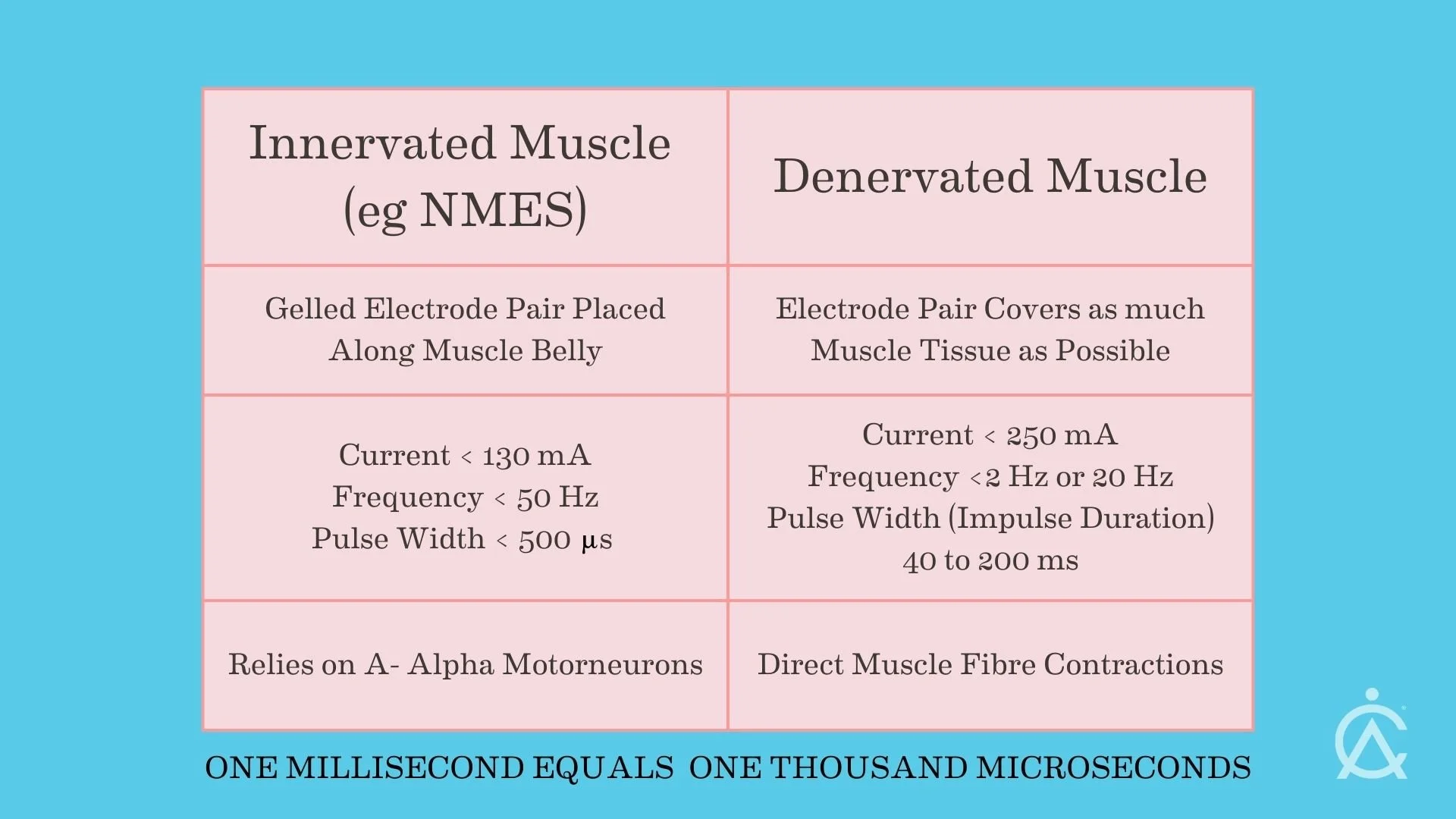
The work of Helmut Kern in Vienna, Ugo Carraro in Padua and their collaborators in the EU RISE project overturned that. The technical key was pulse duration. A healthy nerve responds to pulses lasting well under a millisecond. A denervated muscle fibre, stimulated directly without a nerve to amplify the signal, needs pulses of 120 to 150 milliseconds or more to begin with, hundreds of times longer, delivered at high energy through large electrodes laid over the whole muscle. This is why an ordinary NMES or TENS unit does nothing whatsoever for a denervated muscle, a point I expand on in Normal vs Denervated Muscle. Purpose-built devices are required.
What did the Vienna programme demonstrate? Twenty people with complete, permanent denervation of the thigh muscles from conus and cauda equina injuries completed two years of home-based stimulation, five days a week. Quadriceps cross-sectional area increased by 35%. The average diameter of individual muscle fibres increased by 75%. The force the stimulated muscle could produce increased from 0.8 to 10.3 newton metres, a better than twelvefold rise from an admittedly tiny baseline. A quarter of the participants regained enough force to perform supported stand-up exercises.
For this article, though, the composition data matters most, because this is where "muscle quality" stops being an abstraction. In biopsies of long-term denervated muscle before training, muscle fibre occupied about 26% of the tissue area, connective tissue 62% and fat 13%. After training, muscle fibre occupied 94%, connective tissue under 4% and fat around 2%. Companion imaging work using quantitative CT told the same story at whole-muscle scale: the proportion of the muscle classified as normal tissue rose from about 45% to 60% of its volume, while connective tissue fell by roughly 30% and fat by roughly half. And at the finest scale, electron microscopy showed that 90% or more of the fibres examined had rebuilt their internal contractile architecture and calcium-handling systems, in tissue with no nerve supply of any kind. To my knowledge, this reversal of fatty and fibrous degeneration is the single strongest tissue-quality result anywhere in the electrical stimulation literature.
Nor is this confined to the legs, or to one research group. A Swiss study applied the same principle to denervated hand and forearm muscles in 22 people with tetraplegia: after twelve weeks, ultrasound showed the first dorsal interosseous muscle thickening from 6.3 to 9.2 millimetres, with its internal fibre architecture visibly restored. And in 2025, an American group published the first randomised pilot in this population, finding a 25% increase in rectus femoris cross-sectional area at six months using long-pulse stimulation combined with testosterone treatment. Notably, they also reported that adherence was worse in the long-pulse group than with conventional stimulation, which brings me to the limits.
KEY POINT: Denervated muscle can be rescued, but only with long-pulse stimulation from specialised equipment. Two years of home stimulation, five days a week, restored not just size and force but the composition of the tissue itself, with fat and fibrous tissue retreating from three quarters of the biopsy area to under 6%. No conventional NMES or TENS device can do this.
What the denervation research did not show
I hold the Vienna work in high regard, which is exactly why I want to state its boundaries as clearly as its achievements. This work inspired us to get involved and now we have many clients using the RISE Stimulator from Dr. Schuhfried Medizintechnik GmbH, which evolved from the original RISE research programme.
We should say that the RISE Study outcomes did not restore walking. The documented functional outcome was that around a quarter of participants could perform supported stand-up exercises. The researchers themselves note that only younger and lighter patients can realistically expect to stand and take a few steps with support in parallel bars.
It did not restore normal excitability. Even after two years of successful training, the muscle never regained the electrical responsiveness of healthy innervated tissue. The rebuilt muscle remains a tissue that must be driven by relatively long pulses; it does not become an ordinary muscle again.
It did not involve a randomised comparison. The RISE study was a prospective cohort in which participants served as their own controls over two years. That is respectable evidence, and the tissue-level findings are hard to explain any other way, but a randomised trial against no treatment has never been done, and the only randomised data so far is a ten-person pilot comparing two active treatments.
It did not repair nerves. Nothing in this research claims stimulation causes reinnervation. Conditioning the muscle and regrowing the nerve are separate matters. What muscle conditioning may do, for those whose nerve is expected to regrow, is preserve the tissue so that a returning nerve finds something worth reconnecting to. We rely on this when using the RISE stimulator with injuries where reinnervation is expected.
And it demanded extraordinary commitment. The protocol was stimulation on five days of every week, building to hours of work, sustained for years. The researchers estimate that rebuilding a quadriceps means restoring 2 to 4 million individual fibres, a project they measure in years rather than weeks. Adherence, not biology, is often the binding constraint, and anyone considering this path should hear that before they start rather than discover it afterwards. We are always upfront with clients that using stimulation for denervation works but takes commitment.
On timing, the group's own advice is that treatment should ideally begin within about five years of injury to expect the two-year results described above, and the earlier the better. That said, the picture for late starters is not as bleak as was once assumed, and I have reviewed that evidence separately in Can I Start Electrical Stimulation Years After My Denervation Injury?.
KEY POINT: The denervation evidence shows rescued tissue, not restored function. No walking, no return of normal excitability, no nerve repair, and no randomised trial against no treatment. The protocol demands commitment on most days of the week, over years, and starting within about five years of injury is what the published results describe.
The practical questions this evidence answers
Let me draw the threads together into the questions people actually bring to us.
"Will stimulation keep my muscle alive while my nerve regrows?" This is the peripheral nerve injury question, and the evidence above is the best answer available. Denervated muscle degenerates on a timescale of years, and long-pulse stimulation has been shown to reverse the tissue changes that define that degeneration. What no study can yet tell you is your individual outcome, which depends on your injury, your diagnosis and your consistency. The essential first step is finding out whether your muscle is partly innervated, fully denervated, or recovering, because that determines which kind of stimulation is even relevant. A nerve conduction study answers this, and I have written about what a nerve conduction study actually tells you.
"I bought a stimulator online and nothing happened. Does that mean stimulation will not work for me?" No. It most likely means one of two things: the device was a TENS unit designed for pain relief rather than muscle work, or your muscle is denervated and needs long pulses that no consumer device can produce. A muscle that fails to respond to a conventional stimulator is giving you a form of diagnostic information, not a verdict.
"How much do I need to do?" More than the marketing suggests. In innervated muscle, weekly training volume is what predicts growth. In denervated muscle, the published protocol is work on five days of every week, over years. Anyone offering meaningful tissue change from a few minutes a week is contradicted by every study cited on this page.
"What happens if I stop?" The gains reverse. In people who completed a year of FES cycling and then stopped, only about a fifth of the muscle gain remained a year later, and in the small MRI study above, the fat that had been pushed out of the muscle returned within a month of stopping. Stimulation is exercise, and it follows the rules of exercise. It's true for all of us; we "use it or lose it".
The UK picture, briefly
A question I am asked constantly is why, if the evidence is as described, these treatments are not simply available on the NHS.
For drop foot after stroke, MS or other central neurological conditions, NICE assessed functional electrical stimulation and concluded the evidence "appears adequate to support the use of this procedure". That wording is a permission rather than a recommendation, it dates from 2009, and provision still varies considerably between areas. For FES cycling, NICE has published only a descriptive briefing on one system, which makes again no recommendation, and there is no NICE guidance at all covering stimulation of denervated muscle. Several NHS commissioning policies I have reviewed state plainly that FES beyond drop foot is not routinely funded, citing the evidence gaps discussed above.
I say this to set expectations because for most of the applications in this article, the realistic route in the UK involves an informed conversation with your own clinical team, and in many cases self-funding or case-managed funding. That makes it all the more important that the decision rests on accurate information about what the evidence does and does not show, which is what this article has tried to provide.
What I would suggest
If this topic is relevant to you or someone you support, here is how I would proceed.
First, with your own clinical team, establish what kind of muscle you are dealing with. Ask whether the nerve supply to the affected muscles is intact, and if there is any doubt, ask about a nerve conduction study with EMG. Ask your physiotherapist or consultant directly: is this muscle innervated, partly innervated, or denervated? Every decision that follows depends on that answer.
Second, if the muscle is innervated, discuss with your therapist whether structured stimulation training against genuine load has a place in your programme, and what weekly volume would be realistic for you, since volume is what the evidence rewards.
Where we come in is assessment. At Anatomical Concepts we have spent many years working with both conventional FES, including FES cycling systems, and specialised long-pulse stimulation for denervated muscle, and the starting point is always the same: establishing what your tissue can respond to before any equipment is discussed. Decisions about your treatment belong with you and your clinicians, and nothing we do replaces that relationship.
If you would like to talk any of this through, whether you are at the stage of interpreting a nerve conduction report or weighing up a stimulation system, do get in touch. A conversation costs nothing, and we would rather help you ask the right questions than see you spend money answering the wrong ones.
Further reading
Goodpaster BH, Park SW, Harris TB, et al. The loss of skeletal muscle strength, mass, and quality in older adults: the Health, Aging and Body Composition Study. Journals of Gerontology Series A 2006; 61(10): 1059 to 1064. https://doi.org/10.1093/gerona/61.10.1059
Delmonico MJ, Harris TB, Visser M, et al. Longitudinal study of muscle strength, quality, and adipose tissue infiltration. American Journal of Clinical Nutrition 2009; 90(6): 1579 to 1585. https://doi.org/10.3945/ajcn.2009.28047
Goodpaster BH, Kelley DE, Thaete FL, He J, Ross R. Skeletal muscle attenuation determined by computed tomography is associated with skeletal muscle lipid content. Journal of Applied Physiology 2000; 89(1): 104 to 110. https://doi.org/10.1152/jappl.2000.89.1.104
Young HJ, Jenkins NT, Zhao Q, McCully KK. Measurement of intramuscular fat by muscle echo intensity. Muscle and Nerve 2015; 52(6): 963 to 971. https://doi.org/10.1002/mus.24656
Stock MS, Thompson BJ. Echo intensity as an indicator of skeletal muscle quality: applications, methodology, and future directions. European Journal of Applied Physiology 2021; 121(2): 369 to 380. https://doi.org/10.1007/s00421-020-04556-6
Castro MJ, Apple DF, Hillegass EA, Dudley GA. Influence of complete spinal cord injury on skeletal muscle cross-sectional area within the first 6 months of injury. European Journal of Applied Physiology and Occupational Physiology 1999; 80(4): 373 to 378. https://doi.org/10.1007/s004210050606
Castro MJ, Apple DF, Staron RS, Campos GE, Dudley GA. Influence of complete spinal cord injury on skeletal muscle within 6 mo of injury. Journal of Applied Physiology 1999; 86(1): 350 to 358. https://doi.org/10.1152/jappl.1999.86.1.350
Gorgey AS, Dudley GA. Skeletal muscle atrophy and increased intramuscular fat after incomplete spinal cord injury. Spinal Cord 2007; 45(4): 304 to 309. https://doi.org/10.1038/sj.sc.3101968
Bekhet AH, Jahan AM, Bochkezanian V, et al. Effects of electrical stimulation training on body composition parameters after spinal cord injury: a systematic review. Archives of Physical Medicine and Rehabilitation 2022; 103(6): 1168 to 1178. https://doi.org/10.1016/j.apmr.2021.09.004
Mohr T, Andersen JL, Biering-Sorensen F, et al. Long-term adaptation to electrically induced cycle training in severe spinal cord injured individuals. Spinal Cord 1997; 35(1): 1 to 16. https://doi.org/10.1038/sj.sc.3100343
Ryan TE, Brizendine JT, Backus D, McCully KK. Electrically induced resistance training in individuals with motor complete spinal cord injury. Archives of Physical Medicine and Rehabilitation 2013; 94(11): 2166 to 2173. https://doi.org/10.1016/j.apmr.2013.06.016
Gorgey AS, Khalil RE, Carter W, et al. Skeletal muscle hypertrophy and enhanced mitochondrial bioenergetics following electrical stimulation exercises in spinal cord injury: a randomized clinical trial. European Journal of Applied Physiology 2025; 125(4): 1075 to 1089. https://doi.org/10.1007/s00421-024-05661-6
Crameri RM, Cooper P, Sinclair PJ, Bryant G, Weston A. Effect of load during electrical stimulation training in spinal cord injury. Muscle and Nerve 2004; 29(1): 104 to 111. https://doi.org/10.1002/mus.10522
Mastropietro A, Peruzzo D, Taccogna MG, et al. Multiparametric MRI assessment of morpho-functional muscle changes following a 6-month FES-cycling training program: pilot study in people with a complete spinal cord injury. JMIR Rehabilitation and Assistive Technologies 2025; 12: e64825. https://doi.org/10.2196/64825
Gregory CM, Bickel CS. Recruitment patterns in human skeletal muscle during electrical stimulation. Physical Therapy 2005; 85(4): 358 to 364. https://pubmed.ncbi.nlm.nih.gov/15794706/
Bickel CS, Gregory CM, Dean JC. Motor unit recruitment during neuromuscular electrical stimulation: a critical appraisal. European Journal of Applied Physiology 2011; 111(10): 2399 to 2407. https://doi.org/10.1007/s00421-011-2128-4
Porcari JP, McLean KP, Foster C, et al. Effects of electrical muscle stimulation on body composition, muscle strength, and physical appearance. Journal of Strength and Conditioning Research 2002; 16(2): 165 to 172. https://pubmed.ncbi.nlm.nih.gov/11991766/
Kern H, Carraro U, Adami N, et al. Home-based functional electrical stimulation rescues permanently denervated muscles in paraplegic patients with complete lower motor neuron lesion. Neurorehabilitation and Neural Repair 2010; 24(8): 709 to 721. https://doi.org/10.1177/1545968310366129
Kern H, Boncompagni S, Rossini K, et al. Long-term denervation in humans causes degeneration of both contractile and excitation-contraction coupling apparatus, which is reversible by functional electrical stimulation (FES): a role for myofiber regeneration? Journal of Neuropathology and Experimental Neurology 2004; 63(9): 919 to 931. https://doi.org/10.1093/jnen/63.9.919
Boncompagni S, Kern H, Rossini K, et al. Structural differentiation of skeletal muscle fibers in the absence of innervation in humans. Proceedings of the National Academy of Sciences USA 2007; 104(49): 19339 to 19344. https://doi.org/10.1073/pnas.0709061104
Gargiulo P, Kern H, Carraro U, et al. Quantitative color three-dimensional computer tomography imaging of human long-term denervated muscle. Neurological Research 2010; 32(1): 13 to 19. https://doi.org/10.1179/016164109X12536042424171
Kern H, Hofer C, Modlin M, et al. Denervated muscles in humans: limitations and problems of currently used functional electrical stimulation training protocols. Artificial Organs 2002; 26(3): 216 to 218. https://doi.org/10.1046/j.1525-1594.2002.06933.x
Kern H, Carraro U. Home-based functional electrical stimulation for long-term denervated human muscle: history, basics, results and perspectives of the Vienna rehabilitation strategy. European Journal of Translational Myology 2014; 24(1): 3296. https://doi.org/10.4081/ejtm.2014.3296
Kern H, Carraro U. Home-based functional electrical stimulation of human permanent denervated muscles: a narrative review on diagnostics, managements, results and byproducts revisited 2020. Diagnostics 2020; 10(8): 529. https://doi.org/10.3390/diagnostics10080529
Bersch I, Friden J. Electrical stimulation alters muscle morphological properties in denervated upper limb muscles. EBioMedicine 2021; 74: 103737. https://doi.org/10.1016/j.ebiom.2021.103737
Gorgey AS, Khalil RE, Alazzam A, et al. Testosterone and long-pulse-width stimulation (TLPS) on denervated muscles and cardio-metabolic risk factors after spinal cord injury: a pilot randomized trial. Cells 2025; 14(24): 1974. https://doi.org/10.3390/cells14241974
Burnham R, Martin T, Stein R, et al. Skeletal muscle fibre type transformation following spinal cord injury. Spinal Cord 1997; 35(2): 86 to 91. https://doi.org/10.1038/sj.sc.3100364
Frotzler A, Coupaud S, Perret C, et al. Effect of detraining on bone and muscle tissue in subjects with chronic spinal cord injury after a period of electrically-stimulated cycling: a small cohort study. Journal of Rehabilitation Medicine 2009; 41(4): 282 to 285. https://doi.org/10.2340/16501977-0321
NICE. Functional electrical stimulation for drop foot of central neurological origin. HealthTech guidance HTG178 (formerly IPG278), 2009. https://www.nice.org.uk/guidance/htg178
NICE. RT300 for spinal cord injury rehabilitation. Medtech innovation briefing MIB169, 2019. https://www.nice.org.uk/advice/mib169
Related articles on our site
Normal vs Denervated Muscle: Why the Rules of Electrical Stimulation Change After Nerve Injury
How Muscle Fibre Composition Impacts Electrical Stimulation Effectiveness
Can I Start Electrical Stimulation Years After My Denervation Injury? What the Research Shows
Peripheral Nerve Injury and Electrical Stimulation: Which of Three Situations Are You In?
What a Nerve Conduction Study Actually Tells You, and Why It Matters Before Electrical Stimulation
Articles on this site are researched and drafted with the help of AI tools, then checked, edited and approved by me. Every citation is verified against the original source before publication.