
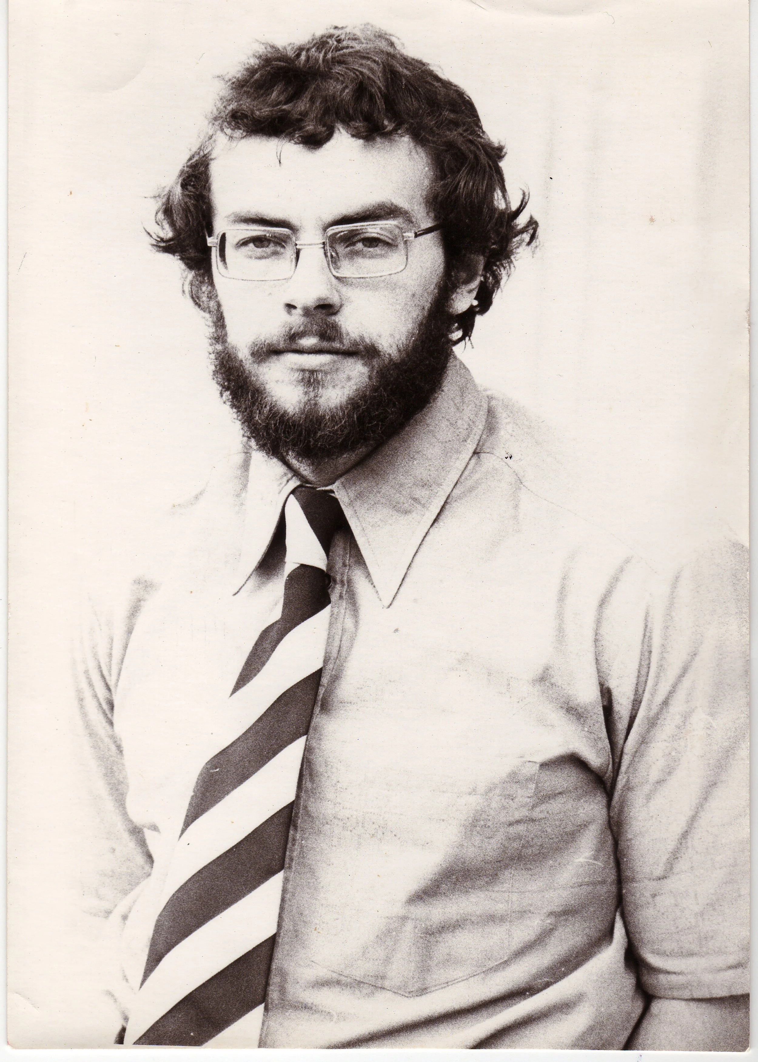
Derek Jones in 1975
The first stimulator I worked with, as a PhD student in the late 1970s, filled a good part of a laboratory bench and needed someone who understood it standing beside it. The Stim2Go electrical stimulation device I carry to homes or clinics now is the size of my hand. It can be strapped to a client's thigh, senses when they start to move, and is set up from an app on my phone. Both deliver a pulse of current to a nerve. Almost everything else about them has changed.
That contrast set me thinking about how the field got from one product to the other, and whether the evolution has a shape. I think it does. In my lifetime, I've seen quite a few changes in the field and seen it come in and out of "fashion". At the moment, we're seeing major steps forward in technology, with, to some extent, the field being held back by a lack of clinical education. Over roughly 250 years, therapeutic electrical stimulation has jumped to a new level perhaps seven times, and each jump was made possible by a new technological development. It might have been a way of handling current, storing it, generating it, shaping it, measuring it, timing it, or using it to tune the nervous system rather than drive it, and finally today triggering it from the person's own movement or intention. These are the stages I allude to in the title.
A second pattern runs alongside the technology, and it is the more useful one for anybody deciding whether to spend money on a stimulator today. At every stage, the clinical claim arrived larger than the evidence, and it took the next twenty years or so to trim it back to size. I think even today, we have a far-from-complete understanding of how best to apply electrical stimulation; sometimes we get results clinically without having any formal understanding of why. The first person to publish on electrical treatment of paralysis noticed this in 1757 and said so. It has happened at every stage since, and it is happening now.
I am not a historian, and I have leaned on the scholarly reviews listed at the end rather than on the folklore, some of which turns out to be wrong.
Stage one: storing a shock
The oldest recorded electrical treatment used a living battery. Scribonius Largus, physician to the emperor Claudius, recommended in the first century that a live torpedo ray be placed on the head of someone with a persistent headache, or under the feet of a person with gout, and held there until the limb went numb. For the next 1,700 years that was more or less the state of the art: a shock you could not dose, from an animal you had to catch.
The first stage in my metaphor, came in 1745 and 1746 with the Leyden jar, a glass vessel that could store charge from a friction machine and release it on command. For the first time a physician could deliver a shock deliberately, to a chosen part of the body, and repeat it. Christian Gottlieb Kratzenstein published what is usually regarded as the first monograph on medical electricity in 1744, and by around 1760 John Wesley, the founder of Methodism, was promoting electrical machines for a long list of conditions. Wesley was a preacher enthusing about a machine, not a clinician reporting results.
Benjamin Franklin was the nearest thing to a clinician reporting results. In a letter to John Pringle dated 21 December 1757, and read to the Royal Society the following month, he described treating paralysed limbs with shocks from Leyden jars. He saw an immediate warmth in the limb and a little more movement on the day, and then no improvement after the fifth day. In his words: "I never knew any advantage from electricity in palsies, that was permanent. And how far the apparent, temporary advantage might arise from the exercise in the patients' journey, and coming daily to my house, or from the spirits given by the hope of success, enabling them to exert more strength in moving their limbs, I will not pretend to say."
KEY POINT: The Leyden jar turned electrical treatment from a curiosity into a procedure that could be dosed and repeated. The first careful report of using it for paralysis, Franklin's in 1757, is also the first report in this field of an immediate effect that did not last, and of an author asking whether it was actually hope and exercise that were doing the work. I do think that electrical stimulation through the ages came to be associated with quackery and fraud and therefore not always taken seriously in a clinical context.
Stage two: generating a current
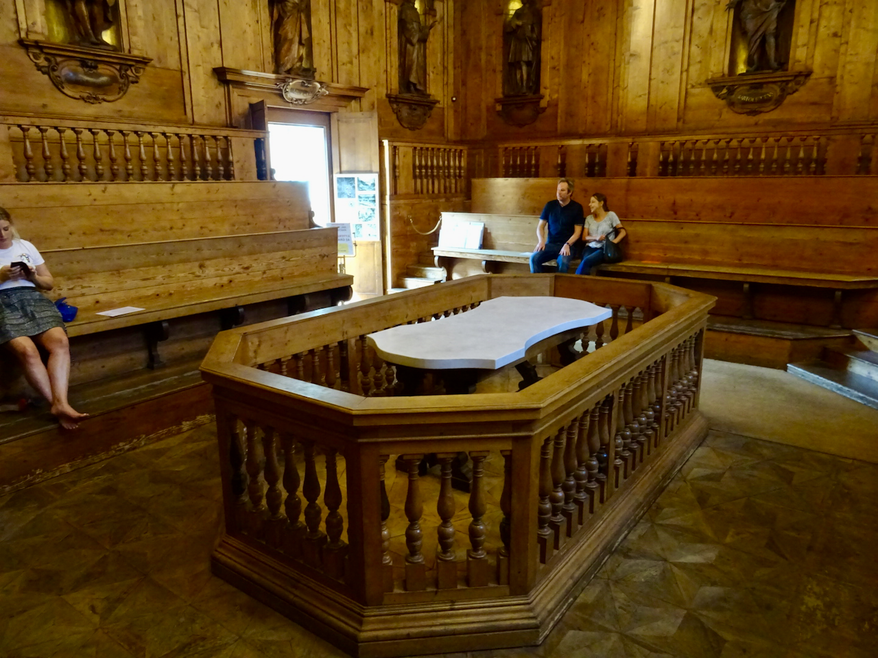
The Anatomical Theatre of the Archiginnasio in Bologna where Galvani would lecture
In 1791 Luigi Galvani, professor of anatomy at Bologna, published his observation that a dissected frog's leg twitched when its nerve was touched by a metal arc, and proposed that living tissue contained its own "animal electricity". Alessandro Volta, professor of physics at Pavia, repeated the experiments, at first agreed, and then argued that the contraction came from the contact between two different metals in the arc, not from anything inside the frog. To prove it he stacked discs of zinc and copper separated by brine-soaked cloth, and in 1800 announced the voltaic pile: the first source of continuous, controllable current.
This is probably the largest single step in the whole story. A Leyden jar gives you a "bang" of energy but no fine control. A pile gives you a tap you can leave running, and everything that follows, from galvanic testing of muscle to the pacemaker, depends on more controllable and sustained current.
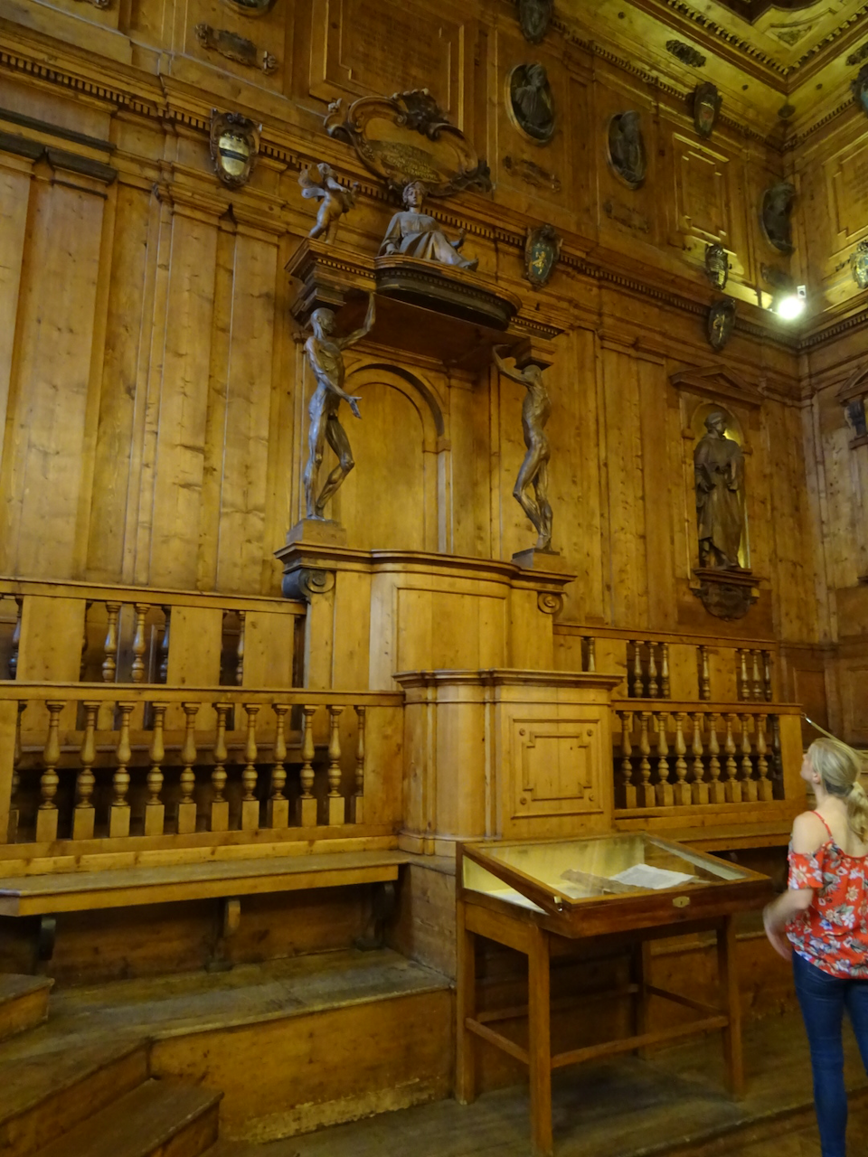
The Spellati (literally meaning "The Skinned Men") are the most famous sculptures inside the Anatomical Theatre of Bologna.
Two corrections to the popular version. Both men were partly right: Volta was correct about the metals, and Galvani was correct that living tissue has its own electrical activity, which he went on to show in preparations with no metal at all and which is the foundation of electrophysiology.
Galvani's nephew Giovanni Aldini took "galvanism" on tour, and in January 1803 applied current to the body of a hanged man at Newgate in front of an audience. Fifteen years later Andrew Ure and James Jeffray did the same in the anatomy theatre at Glasgow University, where I later studied, and I have told that story elsewhere on this site. It is sometimes told as the inspiration for Frankenstein, which is probably a stretch of the imagination. Mary Shelley's novel was published in January 1818 and the Glasgow experiment took place that November. The University's own archive describes the reanimation reading as a myth, and a good example of a claim outrunning its evidence within weeks of the event.
Stage three: shaping the current
A steady current will not make a healthy muscle contract and hold. What makes muscle work is a current that switches on and off, and the technology for producing that reliably arrived with Michael Faraday's discovery of electromagnetic induction in 1831 and the induction coils built on it. The interrupted current they produced was called "faradic", and it is the ancestor of every pulse train a modern stimulator delivers.
The person who worked out what to do with it was Guillaume-Benjamin Duchenne, a physician from Boulogne working in Paris. He is perhaps best known for his name being linked with a form of muscular dystrophy. His treatise on localised electrisation, published in 1855, described stimulating individual muscles through intact skin using moistened surface electrodes in place of the needles then in use. With a faradic current and a well-placed electrode he could make one muscle contract and not its neighbour, and over thirty years he mapped muscle function across the body, including, in his 1862 atlas, the muscles of facial expression. Every self-adhesive electrode pad sold today descends from Duchenne's moist sponge.
Duchenne also noticed that a muscle whose nerve had been damaged responded differently, and the neurologist Wilhelm Erb turned that observation into a bedside test. Between 1867 and 1868 Erb systematised what he called the reaction of degeneration: a denervated muscle loses its response to faradic current altogether, while its response to galvanic current is preserved but changed, with a sluggish, worm-like contraction. Erb credited earlier observers, and the recent historical review by Tatu and Péréon gives priority to Baierlacher, Neumann and Ziemssen. The importance for this site is direct. The reaction of degeneration was the first tool that could separate a muscle with a working nerve from one without, and that distinction still decides whether a client needs a conventional stimulator or the long-pulse equipment used for denervated muscle.
KEY POINT: Faraday's induction coil gave clinicians a repeatable interrupted current, Duchenne showed it could be aimed selectively at one muscle through the skin, and Erb used it to tell an innervated muscle from a denervated one at the bedside. Pulsed current, surface electrodes and the innervated versus denervated distinction are still the working basis of everything we do.
The interlude: when the belt-sellers took over
The same word, electricity, covered a professor mapping facial muscles and a mail-order belt containing a bag of zinc, and by the 1880s the belt was winning. Galvanic chains, electric corsets and the portable "medical battery" were sold directly to the public for nervous debility, impotence, rheumatism and anything else that would sell. Golding Bird's efforts to make electrotherapy respectable at Guy's Hospital, described by the historian Iwan Morus, were swamped by the trade in machines, and by 1912 the American Medical Association's compilation of quackery gave electrical devices a good deal of space.
The profession's response was to retreat into what could be measured. Electrodiagnosis flourished but electrical treatment stalled for half a century. I understand the reflex. Many years ago I was accused, to my face, of being a snake-oil salesman for working with electrical stimulation devices. The accusation drew on a reputation earned in this period and never fully shaken off. The remedy, then and now, is the same: publish the evidence and state its limits.
Stage four: measuring excitability
The fourth stage was not a new way of delivering current but a new way of measuring what current does. In 1909 the French physiologist Louis Lapicque defined two quantities every specialist working with electrical stimulation still uses. These terms are rheobase and chronaxie.
Rheobase is the smallest current that will excite a tissue if left on indefinitely. Chronaxie is the pulse duration needed to excite it at twice that current. Plot current against pulse duration and you have the strength-duration curve, which explains in one picture why a nerve responds to a pulse a few hundred microseconds long while a denervated muscle fibre, with no nerve to amplify the signal, needs a pulse a hundred times longer.
Lapicque then built a theory based on his measurement, proposing that transmission between nerve and muscle required matched chronaxies, but the theory did not survive the physiology that followed. Edgar Adrian's recordings of single nerve fibres in the 1920s, made possible by the valve amplifier and the oscilloscope, replaced it with the picture of nerve impulses we use today. The measurement outlived the explanation. In my experience that is often the fate of a good instrument in this field, and worth remembering when a new device arrives with a new theory attached.
The same period produced the first serious experiment on stimulating denervated muscle. In 1944 Ernst Gutmann and Ludwig Guttmann, the latter at the spinal injuries unit at Stoke Mandeville, reported that galvanic exercise slowed the wasting of denervated muscle in rabbits. The finding that mattered was the dose of application. Stimulation had to be given daily, last at least twenty minutes and produce a vigorous contraction, or it did nothing. That is an animal study, and the human confirmation was still sixty years away, but the dose lesson has held.
KEY POINT: Lapicque's strength-duration curve gave the field a way to measure excitability instead of describing it in vague terms, and it still explains why denervated muscle needs long pulses from specialised equipment. His theory was wrong (as theories often are) but his measurement survived. Gutmann and Guttmann's 1944 rabbit work established that with denervated muscle the dose, not the mere presence of stimulation, decides the result.
A branch line: stimulating to suppress pain
For most people not in our field, electrical stimulation means a TENS machine, so its origin deserves a paragraph even though it branches away from the story of stimulation producing movement. In 1965 Ronald Melzack and Patrick Wall proposed the gate control theory of pain, suggesting that activity in large sensory fibres could close a "gate" in the spinal cord to painful signals. Two years later Norman Shealy and colleagues implanted electrodes on the spinal cord to test the idea, and transcutaneous stimulation, TENS, began partly as a way of screening candidates for the implant before becoming a treatment in its own right. The gate control model has been substantially revised since, and the evidence for TENS has been argued over for sixty years. The stimulation outlived the theory, which by now will sound familiar. I have written about pain applications separately and will leave the branch there.
Stage five: timing the current to a function
Ask anyone in the field of my age when functional electrical stimulation began and they will say 1961, when W. T. Liberson and colleagues published a portable stimulator that fired the peroneal nerve. It is quite common for people with hemiplegia to have problems clearing the foot from the floor to initiate the swing phase of gait. By triggering peroneal nerve stimulation with a carefully positioned foot switch, the foot could be lifted at the correct time to allow a less hesitant gait. The paper's title contains the word "functional", and it was the first time stimulation had been used not to test a muscle, exercise it or relieve pain, but to produce a useful movement at the moment it was needed.
I would put the real pivot point three years earlier and in a different specialty. On 8 October 1958 Åke Senning and Rune Elmqvist implanted the first cardiac pacemaker in Stockholm. That device, a transistorised pulse generator sealed inside the body, is the ancestor of every implanted stimulator in this story, and the transistor is what made Liberson's box portable. Timing stimulation to a functional event was a conceptual step. The technology to do it anywhere outside of a laboratory was borrowed from cardiology.
What followed was thirty years of ambition. In Ljubljana, Alojz Kralj and Tadej Bajd used multichannel surface stimulation to restore standing and a form of walking in paraplegia. In Cleveland, Hunter Peckham's group implanted electrodes in the forearm and hand to restore grasp in tetraplegia, and their Freehand system reached the US market in 1997 after a multicentre trial. The Parastep walking system was approved in 1994. In London, Giles Brindley's sacral anterior root stimulator, a British implant that restored bladder emptying in complete spinal cord injury, was in routine use by the mid-1980s, with the first fifty cases published in 1986. That is a UK first that rarely gets mentioned.
The attempt to create walking-support systems taught the field an uncomfortable lesson. Parastep worked, in the sense that people stood and took steps, but was largely abandoned by its users because walking on electrically driven quadriceps with a rolling frame costs an enormous amount of energy for a short distance. A wheelchair was the more efficient mobility device. Freehand was clinically effective, published, and then commercially withdrawn. The systems that promised walking delivered exercise, and effectiveness did not save them.
The counter-example is British. At Salisbury District Hospital, the Odstock Dropped Foot Stimulator was developed. This was a single-channel device in the Liberson mould, evaluated in a randomised trial in 1997 followed by a five-year clinical audit. NICE assessed the evidence in 2009 and concluded it was adequate to support use with normal clinical governance, in guidance originally numbered IPG278 and now HTG178. The device is still in clinical service three decades on, and I think the reason is that it promised just one thing at one moment in the gait cycle and delivered it.
KEY POINT: The transistor gave birth to the 1958 pacemaker and let stimulation leave the laboratory. Liberson's 1961 foot-drop device was the first to link stimulation to a useful functional activity. The walking systems that followed worked but were abandoned because the energy cost was too high. The single-channel foot-drop stimulator survived because its promise was small and kept.
The "FES bicycle"
FES cycling belongs inside this stage rather than as one of its own, because it changed the purpose of stimulation rather than its fundamental technology. In 1983 Jerrold Petrofsky's group in Ohio described an outdoor tricycle pedalled by stimulated leg muscles, and then in 1984 a stationary ergometer whose stimulator read the crank angle and switched channels against it. The title of the 1984 paper contains the phrase "closed-loop control", which is worth remembering the next time a brochure presents closed-loop cycling as new. Using a bicycle constrained the movement so that the stimulator only had to decide when to apply stimulation to the muscles. This was a much more practical proposition than trying to support the restoration of gait.
Cycling turned FES from a mobility aid into an exercise modality, and the research that followed was about fitness, maintaining bone density and muscle quality, with Ken Hunt's group in Glasgow and Nick Donaldson's in London among the leaders. Angela Frotzler and colleagues found in 2008 that a year of high-volume cycling partially reversed bone loss around the knee in chronic spinal cord injury, and reported the following year that the gains were lost when training stopped. Volume and continuation matter more than the fact of the intervention. At Anatomical Concepts, we have enjoyed more than 20 years of working with FES cycling systems. We were introduced to this area by Ken Hunt and David Allan, as I've written about elsewhere, and they introduced us to Hasomed GmbH, our long-standing partners in stimulation and much more.
FES cycling as offered today requires muscles with an intact nerve supply. Those clients with denervated muscle require different interventions. The Vienna group led by Helmut Kern showed in 1999 that people with long-standing complete denervation could stand with the help of very long, high-charge pulses applied directly to the muscle, and their later home-based programme restored muscle structure over two years. Today, we work with many clients using the RISE stimulator to support persons with peripheral nerve injuries or spinal cord injuries resulting in damage to the lower motor neurons. That is Lapicque's curve put to work, and in fact, the RISE Stimulator from Schuhfried GmbH has an impulse testing function built in to allow the characteristic strength duration curve to be established.
Stage six: modulating rather than driving
The sixth stage was not a new stimulator in my view. It was a new map. Through the 1990s and 2000s, work in animals and then in humans showed that the circuitry in the lumbar spinal cord retains the machinery for standing and stepping after a spinal cord injury, and that a steady "background of stimulation" can bring it close enough to threshold for whatever descending signal survives to become usable. The stimulator stopped being just the sole engine of the movement and became a volume control on the person's own attempt.
The precedent was in the brain, where Alim-Louis Benabid's 1987 report of high-frequency thalamic stimulation for tremor showed that a well-placed electrode could quieten a circuit rather than amplify it. In the spinal cord, Susan Harkema and colleagues reported in 2011 that epidural stimulation allowed a man with a motor-complete injury to stand and move his legs voluntarily while the stimulator was on. Claudia Angeli's group reported over-ground stepping in 2018, and Fabien Wagner's group in Lausanne reported walking with stimulation timed to the intended phase of gait the same year.
I want to be precise about numbers, because the headlines that tend to be in the public eye were not. Those landmark papers together describe fewer than a dozen participants, every one of whom needed the stimulator on, intensive supervised training, and a walker or body-weight support. Two of the four people in the 2018 study stepped over ground with assistance. That is a remarkable result, but it is not yet "paralysed man walks again".
The non-invasive version, transcutaneous spinal cord stimulation through electrodes on the skin over the spine, is the form my clients are most likely to meet, and I have written about it in detail elsewhere on this site.
The largest trial, Up-LIFT, reported in 2024 that stimulation combined with training improved arm and hand function in chronic tetraplegia, and led to US authorisation of the ARC-EX device in December 2024. It was a single-arm trial with no sham comparison, so improvement against baseline cannot be attributed to the stimulation alone. A separate randomised, sham-controlled trial of transcutaneous stimulation for spasticity in multiple sclerosis, reported in 2025 by a group that includes the engineers behind the stimulator we supply (Stim2Go), did not meet its primary endpoint. Both results belong in any fair account.
The strongest evidence in this section comes from a different nerve. In 2021 Jesse Dawson and colleagues reported a randomised, blinded, sham-controlled trial in which vagus nerve stimulation paired with arm rehabilitation after stroke produced more improvement than rehabilitation with sham stimulation. It happens to be the one intervention here that requires an implant and a surgeon, which says something about what good evidence costs. However, we have recently agreed to work with a tVNS device from Germany, and we have a new website at tvns-uk.com that introduces the technology. I have long had an interest in the vagus nerve because of its fundamental role in autonomic nervous system function. This device accesses the vagus nerve with a custom electrode placed in the left ear, so no surgical intervention is needed. Applying stimulation this way is not a quick fix, and we are keen to explore the value of tVNS in rehabilitation.
KEY POINT: Since the late 1980s stimulation has increasingly been used to raise the excitability of surviving circuits so that a person's own effort becomes effective, rather than to produce the movement itself. The results are real but the participant numbers are small. The best-evidenced application, paired vagus nerve stimulation for stroke, is randomised and sham-controlled; the most talked-about, transcutaneous spinal stimulation, is not yet.
Stage seven: sensing the person
The seventh stage is the one we are in, and the reason I began with the Stim2Go device on my client's thigh. Sensors have become cheap, small and reliable. Accelerometers and gyroscopes cost pence, a microcontroller can run a control loop from a coin-sized battery, and the stimulator has stopped being the most interesting part of the system. What is interesting now is what tells it to fire. We are in an age where modern control systems become the way we refine outcomes.
My original degree was in the science of control systems and one of the things we learned very quickly is that if we want to control something, first we need to be able to measure its status over time. There are, in a simplistic sense, two types of control system: open-loop and closed-loop. For most of the history of electrical stimulation, we've been working with open-loop systems. For example, we apply energy to the body, a muscle contracts, and we get an outcome as a limb movement. However, with an open-loop system, at the input where we apply the energy, there is no information available about the outcome. We are hoping there is a rational relationship between the energy we apply and the outcome we get. With a closed-loop system, there is for example, deliberate measurement of the state of the outcome, which is fed back to a functional component that can compare the desired outcome with the actual outcome in real time. The difference between the desired outcome and the present outcome can be used to drive the system. That is closed-loop control.
The idea of using a closed-loop control system is older than the hardware. In 1998 Gerard Francisco and colleagues reported a randomised pilot in which stimulation of the wrist extensors after stroke was triggered when the patient's own attempt produced a detectable EMG signal. In this system, every stimulated contraction was paired with an intention. Reviews were sceptical for a decade, and a 2019 meta-analysis by Kátia Monte-Silva's group found a benefit for hand and wrist recovery, with the usual caveats about small trials and difficult blinding. The claim that triggered stimulation beats stimulation on a timer is plausible, supported by what we see in practice, but it is still under-evidenced for the purist.
Some of what is happening now is genuinely new. Reading a person's "intention" from the motor cortex and using it to trigger muscle stimulation was first done in a person by Chad Bouton's group in 2016, followed by Bolu Ajiboye's group in 2017, and in 2023 Henri Lorach and colleagues connected a cortical implant to a spinal stimulator so that a man with a spinal cord injury could walk with his own intention setting the pace. Continuous, real-time, estimation of gait phase from an inertial sensor has replaced Liberson's heel switch.
Some of it is old technology in new packaging. EMG triggering dates from 1998. Closed-loop control of cycling is in the title of a 1984 paper. "Personalised parameters" usually means setting the current to the individual, which Duchenne did by hand in 1855. When a device is described as intelligent or adaptive, the question to ask is which of these it means.
And the evidence is thin in exactly the places the excitement is greatest. The three cortical papers describe three participants, one each: engineering achievements of a high order, and not yet clinical evidence. For the more ordinary devices, the most useful finding for a UK reader is also the least flattering to the industry: Sarah Prenton's meta-analysis found that a foot-drop stimulator and a conventional ankle brace produce broadly similar effects on walking. The case for the stimulator rests on personal preference, secondary effects and ease of long-term use, not on superior walking speed.

The Stim2Go NMES unit
The Stim2Go device on my client's thigh sits in this landscape. Its motion sensing lets stimulation wait for the person's own attempt to stand, pedal or reach, on whatever equipment they already own, and I have described the practical side of that in separate articles. The loop it closes is the one Petrofsky closed in 1984. What is new is the freedom to close it anywhere and control it all from an app, which is a significant practical advance rather than a purely scientific one.
KEY POINT: The availability of tiny, inexpensive sensors makes it possible to trigger stimulation from a person's own movement or intention, which pairs every contraction with an attempt and is the most promising direction in the field. Cortical control has been shown in single individuals but is not yet at the level of clinical evidence. Much of what is marketed as new dates from the 1980s and 1990s as concept, and a foot-drop stimulator does not let you walk faster than a brace.
The pattern, and how to read the next claim
Seven stages, then, and the same mechanism at each. A new way of handling current, from the Leyden jar to the accelerometer, lets the field act at a new level. The clinical claim arrives immediately and is generally wider than the evidence can support initially. Over the next twenty years the claim is trimmed, the instrument survives and improves, and whatever theory came attached to it is usually replaced. Franklin's letter of 1757 would serve as a summary of most of what followed.
None of this is an argument against electrical stimulation. I have spent a significant part of my working life on it, and the trimmed-back claims are still substantial: a foot lifted at the right moment, a bladder emptied, a paralysed muscle kept alive for the years a nerve takes to regrow. It is an argument for reading each new claim with the pattern in mind, and asking four questions. What is the enabling technology, and is it actually new? How many participants were studied? Was there a sham or control condition? And what happened when the training stopped?
Conclusion
Applying electricity to the body for therapeutic purposes has a very long history. The root of this is that applying energy to the body can produce a physiological effect, which we hope has a therapeutic benefit. It's always been easier to develop and apply the technology than it is to understand its consequences. We are in an era now where closed-loop control is enabling much more fine control of the consequences of stimulation. We expect this approach to be more supportive of functional recovery and rehabilitation, but we can expect that the road to evidence will be a long and difficult one at times. To some extent, I see that many clinicians today still have very little knowledge of electrical stimulation and its variants. Are we still being held back by the perception that, once upon a time, this was all about quackery? As an engineer, I'm always interested in how to apply technology to medicine and healthcare. It was that curiosity that took me into the field of bioengineering in the first place. However, innovation and the development and application of technology have, at some point, to be tied into the clinical evidence of benefit. As the medical device regulations become more challenging to respond to, my fear is that we won't explore technology to take things to the new level. You might argue that we should work with the tools that we have right now and strive to get a better evidence base for these.
At Anatomical Concepts we have worked with functional electrical stimulation, FES cycling, transcutaneous spinal cord stimulation and long-pulse stimulation for denervated muscle over many years, and the starting point is always the same: establishing what your nervous system and muscles can respond to before any equipment is discussed. Decisions about your treatment belong with you and your clinicians, and nothing we do replaces that relationship.
If you would like to talk any of this through, whether you are trying to make sense of a claim you have read or deciding between two approaches, do get in touch. A conversation costs nothing, and I would rather help you ask the right questions than see you spend money answering the wrong ones.
Further reading
Kane K, Taub A. A history of local electrical analgesia. Pain 1975; 1(2): 125 to 138. https://doi.org/10.1016/0304-3959(75)90097-4
Tsoucalas G, Karamanou M, Lymperi M, Gennimata V, Androutsos G. The "torpedo" effect in medicine. International Maritime Health 2014; 65(2): 65 to 67. https://doi.org/10.5603/IMH.2014.0015
Kaplan PW. The real Dr Frankenstein: Christian Gottlieb Kratzenstein? Journal of the Royal Society of Medicine 2002; 95(11): 577 to 578. https://doi.org/10.1258/jrsm.95.11.577
Franklin B. Letter to John Pringle, 21 December 1757, on the effects of electricity in paralytic cases. Royal Society archive L&P/3/278 (https://makingscience.royalsociety.org/items/l-and-p_3_278/letter-of-electricity-in-paralytic-cases-from-benjamin-franklin-to-john-pringle). Text in: Morse RA (ed). Benjamin Franklin: Papers on Electricity. 2004; pages 206 to 208. https://psrc.aapt.org/Franklin/pdf/franklin_electricity_print.pdf
Heidland A, Fazeli G, Klassen A, et al. Neuromuscular electrostimulation techniques: historical aspects and current possibilities in treatment of pain and muscle waisting [sic]. Clinical Nephrology 2013; 79 Suppl 1: S12 to S23. https://pubmed.ncbi.nlm.nih.gov/23249528/
Piccolino M. Luigi Galvani and animal electricity: two centuries after the foundation of electrophysiology. Trends in Neurosciences 1997; 20(10): 443 to 448. https://doi.org/10.1016/s0166-2236(97)01101-6
Piccolino M. Animal electricity and the birth of electrophysiology: the legacy of Luigi Galvani. Brain Research Bulletin 1998; 46(5): 381 to 407. https://doi.org/10.1016/s0361-9230(98)00026-4
Parent A. Giovanni Aldini: from animal electricity to human brain stimulation. Canadian Journal of Neurological Sciences 2004; 31(4): 576 to 584. https://doi.org/10.1017/s0317167100003851
University of Glasgow. Andrew Ure. The University of Glasgow Story, accessed 3 September 2026. https://universitystory.gla.ac.uk/people/WH17224
Valenstein ES. The discovery of chemical neurotransmitters. Brain and Cognition 2002; 49(1): 73 to 95. https://doi.org/10.1006/brcg.2001.1487
Tatu L, Péréon Y. The origins of neuromuscular electrodiagnosis, 1800 to 1950: a crucial period. European Neurology 2025; 88(1): 32 to 39. https://doi.org/10.1159/000544957
Parent A. Duchenne de Boulogne: a pioneer in neurology and medical photography. Canadian Journal of Neurological Sciences 2005; 32(3): 369 to 377. https://doi.org/10.1017/s0317167100004315
Morus IR. Marketing the machine: the construction of electrotherapeutics as viable medicine in early Victorian England. Medical History 1992; 36(1): 34 to 52. https://doi.org/10.1017/s0025727300054612
Wexler A. The medical battery in the United States (1870 to 1920): electrotherapy at home and in the clinic. Journal of the History of Medicine and Allied Sciences 2017; 72(2): 166 to 192. https://doi.org/10.1093/jhmas/jrx001
Irnich W. The terms "chronaxie" and "rheobase" are 100 years old. Pacing and Clinical Electrophysiology 2010; 33(4): 491 to 496. https://doi.org/10.1111/j.1540-8159.2009.02666.x
Gutmann E, Guttmann L. The effect of galvanic exercise on denervated and re-innervated muscles in the rabbit. Journal of Neurology and Psychiatry 1944; 7(1-2): 7 to 17. https://doi.org/10.1136/jnnp.7.1-2.7
Melzack R, Wall PD. Pain mechanisms: a new theory. Science 1965; 150(3699): 971 to 979. https://doi.org/10.1126/science.150.3699.971
Shealy CN, Mortimer JT, Reswick JB. Electrical inhibition of pain by stimulation of the dorsal columns: preliminary clinical report. Anesthesia and Analgesia 1967; 46(4): 489 to 491. https://pubmed.ncbi.nlm.nih.gov/4952225/
Liberson WT, Holmquest HJ, Scot D, Dow M. Functional electrotherapy: stimulation of the peroneal nerve synchronized with the swing phase of the gait of hemiplegic patients. Archives of Physical Medicine and Rehabilitation 1961; 42: 101 to 105. https://pubmed.ncbi.nlm.nih.gov/13761879/
Aquilina O. A brief history of cardiac pacing. Images in Paediatric Cardiology 2006; 8(2): 17 to 81. https://pmc.ncbi.nlm.nih.gov/articles/PMC3232561/
Kralj A, Bajd T, Turk R, Krajnik J. Gait restoration in paraplegic patients: a feasibility demonstration using multichannel surface electrode FES. Journal of Rehabilitation Research and Development 1983; 20(1): 3 to 20. https://pubmed.ncbi.nlm.nih.gov/6887064/
Brindley GS, Polkey CE, Rushton DN, Cardozo L. Sacral anterior root stimulators for bladder control in paraplegia: the first 50 cases. Journal of Neurology, Neurosurgery and Psychiatry 1986; 49(10): 1104 to 1114. https://doi.org/10.1136/jnnp.49.10.1104
Peckham PH, Keith MW, Kilgore KL, et al. Efficacy of an implanted neuroprosthesis for restoring hand grasp in tetraplegia: a multicenter study. Archives of Physical Medicine and Rehabilitation 2001; 82(10): 1380 to 1388. https://doi.org/10.1053/apmr.2001.25910
Graupe D, Kohn KH. Functional neuromuscular stimulator for short-distance ambulation by certain thoracic-level spinal-cord-injured paraplegics. Surgical Neurology 1998; 50(3): 202 to 207. https://doi.org/10.1016/s0090-3019(98)00074-3
Burridge JH, Taylor PN, Hagan SA, Wood DE, Swain ID. The effects of common peroneal stimulation on the effort and speed of walking: a randomized controlled trial with chronic hemiplegic patients. Clinical Rehabilitation 1997; 11(3): 201 to 210. https://doi.org/10.1177/026921559701100303
Taylor P, Burridge J, Dunkerley A, et al. Clinical audit of 5 years provision of the Odstock dropped foot stimulator. Artificial Organs 1999; 23(5): 440 to 442. https://doi.org/10.1046/j.1525-1594.1999.06374.x
NICE. Functional electrical stimulation for drop foot of central neurological origin. HealthTech guidance HTG178 (formerly IPG278), 2009. https://www.nice.org.uk/guidance/htg178
Petrofsky JS, Heaton H, Phillips CA. Outdoor bicycle for exercise in paraplegics and quadriplegics. Journal of Biomedical Engineering 1983; 5(4): 292 to 296. https://doi.org/10.1016/0141-5425(83)90003-1
Petrofsky JS, Heaton HH, Phillips CA. Leg exerciser for training of paralysed muscle by closed-loop control. Medical and Biological Engineering and Computing 1984; 22(4): 298 to 303. https://doi.org/10.1007/BF02442096
Hunt KJ, Fang J, Saengsuwan J, Grob M, Laubacher M. On the efficiency of FES cycling: a framework and systematic review. Technology and Health Care 2012; 20(5): 395 to 422. https://doi.org/10.3233/THC-2012-0689
Frotzler A, Coupaud S, Perret C, et al. High-volume FES-cycling partially reverses bone loss in people with chronic spinal cord injury. Bone 2008; 43(1): 169 to 176. https://doi.org/10.1016/j.bone.2008.03.004
Frotzler A, Coupaud S, Perret C, et al. Effect of detraining on bone and muscle tissue in subjects with chronic spinal cord injury after a period of electrically-stimulated cycling: a small cohort study. Journal of Rehabilitation Medicine 2009; 41(4): 282 to 285. https://doi.org/10.2340/16501977-0321
Kern H, Hofer C, Strohhofer M, et al. Standing up with denervated muscles in humans using functional electrical stimulation. Artificial Organs 1999; 23(5): 447 to 452. https://doi.org/10.1046/j.1525-1594.1999.06376.x
Kern H, Carraro U, Adami N, et al. Home-based functional electrical stimulation rescues permanently denervated muscles in paraplegic patients with complete lower motor neuron lesion. Neurorehabilitation and Neural Repair 2010; 24(8): 709 to 721. https://doi.org/10.1177/1545968310366129
Benabid AL, Pollak P, Louveau A, Henry S, de Rougemont J. Combined (thalamotomy and stimulation) stereotactic surgery of the VIM thalamic nucleus for bilateral Parkinson disease. Applied Neurophysiology 1987; 50(1-6): 344 to 346. https://doi.org/10.1159/000100803
Harkema S, Gerasimenko Y, Hodes J, et al. Effect of epidural stimulation of the lumbosacral spinal cord on voluntary movement, standing, and assisted stepping after motor complete paraplegia: a case study. Lancet 2011; 377(9781): 1938 to 1947. https://doi.org/10.1016/S0140-6736(11)60547-3
Angeli CA, Boakye M, Morton RA, et al. Recovery of over-ground walking after chronic motor complete spinal cord injury. New England Journal of Medicine 2018; 379(13): 1244 to 1250. https://doi.org/10.1056/NEJMoa1803588
Wagner FB, Mignardot JB, Le Goff-Mignardot CG, et al. Targeted neurotechnology restores walking in humans with spinal cord injury. Nature 2018; 563(7729): 65 to 71. https://doi.org/10.1038/s41586-018-0649-2
Moritz C, Field-Fote EC, Tefertiller C, et al. Non-invasive spinal cord electrical stimulation for arm and hand function in chronic tetraplegia: a safety and efficacy trial. Nature Medicine 2024; 30(5): 1276 to 1283. https://doi.org/10.1038/s41591-024-02940-9
US Food and Drug Administration. De Novo classification request DEN240014, ARC-EX System, decision 19 December 2024. https://www.accessdata.fda.gov/scripts/cdrh/cfdocs/cfpmn/denovo.cfm?id=DEN240014
Spieker EL, Hoffmann M, Otto C, et al. Short-term effect of transcutaneous spinal cord stimulation in patients with multiple sclerosis: a randomized sham-controlled crossover study. Frontiers in Neurology 2025; 16: 1618519. https://doi.org/10.3389/fneur.2025.1618519
Dawson J, Liu CY, Francisco GE, et al. Vagus nerve stimulation paired with rehabilitation for upper limb motor function after ischaemic stroke (VNS-REHAB): a randomised, blinded, pivotal, device trial. Lancet 2021; 397(10284): 1545 to 1553. https://doi.org/10.1016/S0140-6736(21)00475-X
Francisco G, Chae J, Chawla H, et al. Electromyogram-triggered neuromuscular stimulation for improving the arm function of acute stroke survivors: a randomized pilot study. Archives of Physical Medicine and Rehabilitation 1998; 79(5): 570 to 575. https://doi.org/10.1016/s0003-9993(98)90074-0
Monte-Silva K, Piscitelli D, Norouzi-Gheidari N, et al. Electromyogram-related neuromuscular electrical stimulation for restoring wrist and hand movement in poststroke hemiplegia: a systematic review and meta-analysis. Neurorehabilitation and Neural Repair 2019; 33(2): 96 to 111. https://doi.org/10.1177/1545968319826053
Bouton CE, Shaikhouni A, Annetta NV, et al. Restoring cortical control of functional movement in a human with quadriplegia. Nature 2016; 533(7602): 247 to 250. https://doi.org/10.1038/nature17435
Ajiboye AB, Willett FR, Young DR, et al. Restoration of reaching and grasping movements through brain-controlled muscle stimulation in a person with tetraplegia: a proof-of-concept demonstration. Lancet 2017; 389(10081): 1821 to 1830. https://doi.org/10.1016/S0140-6736(17)30601-3
Lorach H, Galvez A, Spagnolo V, et al. Walking naturally after spinal cord injury using a brain-spine interface. Nature 2023; 618(7963): 126 to 133. https://doi.org/10.1038/s41586-023-06094-5
Azevedo Coste C, Jovic J, Pissard-Gibollet R, Froger J. Continuous gait cycle index estimation for electrical stimulation assisted foot drop correction. Journal of NeuroEngineering and Rehabilitation 2014; 11: 118. https://doi.org/10.1186/1743-0003-11-118
Prenton S, Hollands KL, Kenney LP. Functional electrical stimulation versus ankle foot orthoses for foot-drop: a meta-analysis of orthotic effects. Journal of Rehabilitation Medicine 2016; 48(8): 646 to 656. https://doi.org/10.2340/16501977-2136
Related articles on our site
Understanding the Cost of Care: Why Medical-Grade Stimulation Devices Outprice Consumer Units
Transcutaneous Spinal Cord Stimulation: What Patients Need to Know
Spasticity and Muscle Spasms: A Plain English Guide for People Living With Them
Standing Up Again: How Responsive Electrical Stimulation Can Support Sit-to-Stand Practice
Articles on this site are researched and drafted with the help of AI tools, then checked, edited and approved by me. Every citation is verified against the original source before publication.