
Tissue mechanics has been a thread throughout much of my career and not always a welcome one because it's sometimes a reminder of problems that can be frustratingly difficult to deal with. Over decades, healthcare systems have recognised that pressure ulcers represent a significant sink for resources and can be a life threatening challenge for those affected. From time to time we might read that "pressure ulcers are preventable" or that they represent a "failure of nursing". I think such statements can be unhelpful as they hint at prevention being simpler than it actually is in many cases. Let me elaborate.
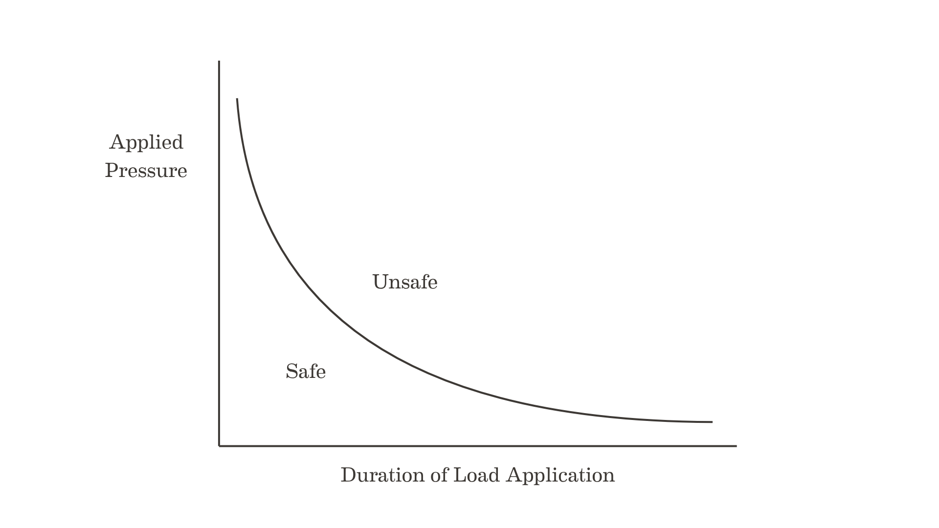
A stylised representation of the effect of pressure and load application time on tissue viability
Back in the 1970s Reswick and Rogers published their study in Bed Sore Biomechanics that attempted to map the pressure tolerance of human tissues. It's a diagram I still see used in education today. It displays the relationship between applied pressure in mmHg and time of pressure application, with combinations of pressure and time either falling in a safe region or carrying a risk of ulceration. It suggests that even high pressures can be safe if only applied for a short period of time.
Whilst this might contain a grain of truth it is very situation dependent, an oversimplification and potentially dangerous to rely on in practice.
As a career I have found engineering applied to medicine (Bioengineering) to be an extremely rewarding and challenging endeavour. The body is a miraculous machine with layers of complexity that are not always obvious until you seek to interact with it. During my career I was fortunate to spend time with the late James (Jim) Foort who, along with Chuck Radcliffe at the University of California had developed the patellar-tendon-bearing (PTB) lower limb prosthesis. Their concept was a significant improvement for amputees and created by changing the nature of the body-device interface that controls the quality of fit. They remodelled the way the semi-rigid structures of a prosthetic socket interacted with the living tissues of the amputee's stump. They were actively manipulating pressure and shear at the tissue interface. I remember Jim, who entered the field as a smart aeronautical engineer, telling me that when he first looked at the problem he thought he would have it solved in months. In fact, much of his long career was spent trying to understand its complexities.
When we read clinical guidelines for pressure ulcer prevention we will find mention of the terms pressure and shear but usually no real explanation of what these actually mean within the concept of tissue mechanics.
Consider this case of a referral from the community to a diabetic foot clinic. This lady presented one July with a painful and sloughy black heel with a medial ulcer which was worsening despite 6 weeks treatment (lotions and potions) from her GP practice. In September, she returned to the clinic with the wound shown here.
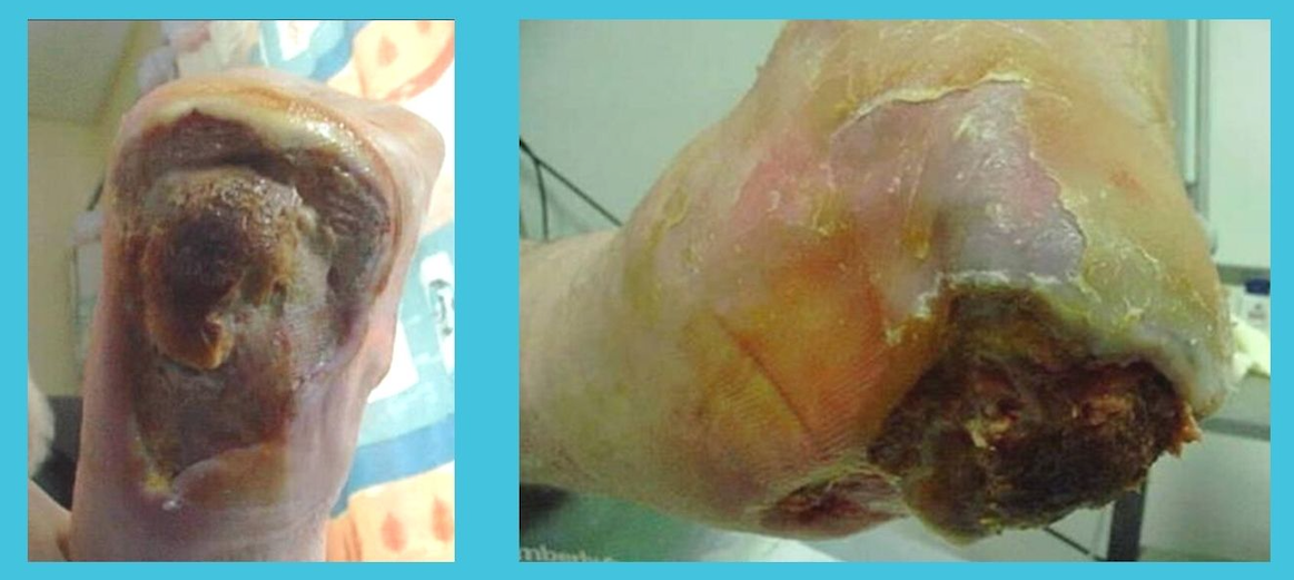
This lady refused amputation despite severe ulceration, neuropathy and infection. This was actually healed with medical interventions and total pressure relief although it took 156 weeks.
In cases such as this with neuropathy, ischaemia and infection it would be difficult to say that even pressure offloading would have been sufficient to prevent this situation as the medical factors were doing much of the damage. However, there is no doubt that the application of total pressure relief was indicated. When pressure ulcers develop there are always mechanical and medical risk factors as we have written about previously and which factors dominate will vary from case to case. It just seems to me sensible to eliminate any risk that we can because the economic and human costs of failure are so high.
I have sat in many meetings where the people using the words pressure and shear had never been shown what they mean in a mechanical sense, and had no way of knowing that the tissue under the skin behaves very differently from the skin itself. The rest of this article explores these terms and uncovers some of the complexity that is to be found when we look beyond the clinical guidance.
The definitions everyone quotes
The current NHS definition, set out by NHS Improvement in 2018, describes a pressure ulcer as localised damage to the skin and underlying tissue, usually over a bony prominence or related to a device, "resulting from sustained pressure (including pressure associated with shear)". The 2025 edition of the International Pressure Injury Guideline says much the same: damage "resulting from prolonged pressure or pressure in combination with shear".
Both definitions are sound. Both also contain three ideas that do all the work and are rarely explained to the people expected to act on them: pressure, shear, and the word "sustained". As we hinted above, it is very difficult to know what is meant by "sustained". What follows is about those three, and about a fourth that neither definition mentions: the tissue doing the responding is soft, layered and time-dependent in ways that make the simple picture misleading.
KEY POINT: Every definition of a pressure ulcer rests on three undefined ideas: pressure, shear and time. Understanding what each one actually does to the tissue under the skin is the difference between following a policy and knowing why it works, and why it sometimes fails.
Pressure is a force spread over an area
A force is a push. Pressure is that push divided by the area it is spread over. The same push concentrated on a small area produces a high pressure; spread over a large area it produces a low one. A drawing pin demonstrates the principle: the thumb pushes on the broad head and feels nothing, the point delivers the identical force to the wall through an area a thousand times smaller, and the wall yields.
Engineers measure pressure in kilopascals (kPa). Clinicians more often meet millimetres of mercury (mmHg), and 1 kPa is about 7.5 mmHg. Older literature is organised around a figure of 32 mmHg, measured in the 1930s in nail-fold capillaries and later treated as the pressure at which capillaries close. It is a historical figure rather than a reliable damage threshold, and the research I will come to has moved well beyond it, though product literature still quotes it as if it is reliable.
The important phrase in the definition is "over a bony prominence". When a body rests on a surface, the load passes through the soft tissue to the skeleton. Where the skeleton comes close to the surface with little tissue in between, the force is concentrated through a small area, and that is where the pressure is highest. The sacrum, the ischial tuberosities, the trochanters and the heels are on the familiar list for areas at risk due to these mechanical factors.
The heel is the extreme case. The calcaneus is a relatively sharp, curved bone, covered posteriorly by a small volume of soft tissue, and the weight of the whole lower leg in a supine patient rests through it on an area of a few square centimetres. The international guideline's heel chapter puts the consequence plainly: given the small heel surface area, redistributing the load is "challenging". That sentence carries most of what follows.
Shear is the sideways force, and friction is what delivers it
Pressure acts at right angles ("normal") to the skin surface, pushing inwards. Shear acts along the skin, parallel to the surface. The textbook definition, and the one the 2022 international aetiology review uses, is that shear stress is the sideways force per unit area, exactly as pressure is the perpendicular force per unit area.
Press the palm flat on a table. Pushing straight down is pressure. Now try to slide the hand along the table while still pressing. The table resists the slide; that resistance is friction, and the hand does not move. But the skin of the palm is dragged slightly in the direction of the attempted slide while the bones stay put, and the tissue between is distorted sideways. That distortion is shear strain, and the force producing it is shear.
At the bedside the classic picture is a patient sliding down a bed whose head has been raised. The skin of the sacrum and heels is held by friction against the sheet, the skeleton slides under gravity, and the tissue in between is stretched. No movement need be visible. Friction is what prevents the skin from moving, and therefore what transmits the shear into the tissue. Moisture matters here for a reason that is often misunderstood: moist skin has a higher coefficient of friction against hospital fabric, not a lower one. In a human study of forearm skin against hospital textile, friction rose by roughly 26 to 43 per cent from very dry to normally moist skin, and more than doubled against fully wet fabric. A damp heel on a sheet grips harder and shears more.
Why does shear matter so much? The reference experiment is nearly fifty years old. In 1979 Bennett and colleagues applied controlled pressure and shear to the thenar eminence (the pad at the base of the thumb) of four healthy volunteers and measured blood flow. Pressure alone was roughly twice as effective as shear alone at reducing flow, but when a moderate level of shear was added, the pressure needed to stop the flow fell by about half. This was a small sample with healthy tissue, and not applied to the heel. The finding has nevertheless held up: shear does not merely add to pressure, it lowers the bar at which pressure does harm.
The current synthesis, set out by Gefen in 2024, is that superficial damage to the skin is driven mainly by shear, while deeper damage under a bony prominence arises from pressure concentrated over the bone combined with shear at the surface. I have written separately about shear at the heel; here I want to stay with the general principle.
KEY POINT: Pressure pushes in; shear drags sideways; friction holds the skin so that shear reaches the tissue. Moist skin grips harder, not less. In the one classic human experiment, adding shear roughly halved the pressure needed to shut off blood flow, which is why the two are always named together.
Stress is what you apply, strain is what the tissue does
Here is the distinction that almost every explanation I have read skips, and that turns out to be the centre of the whole subject. Stress is a load per unit area. Pressure is one kind of stress, the perpendicular kind but shear stress is the other. Both describe what is being applied to the tissue.
Strain is how much the tissue deforms in response. Squash a block of tissue to three-quarters of its height and it has experienced 25 per cent compressive strain. Drag the top sideways relative to the bottom and it has experienced shear strain. Strain describes what is happening inside the material.
The distinction matters because cells are not killed by stress. They are killed by being deformed. A cell stretched or flattened far enough, for long enough, loses the integrity of its membrane and dies, whether or not anyone was measuring the load that did it. The same stress applied to stiff tissue and to soft tissue produces very different strains, and it is the strain that does the damage. This means that whenever tissue is in contact with a support surface that deforms tissue we have an "at-risk" situation.
This is not my private view. In 2010 Oomens, Loerakker and Bader, three of the engineers who have done most to reshape this field, published a paper in the Journal of Tissue Viability with the stated aim of explaining to readers "with no conventional background in mechanics" why interface pressure is not the right parameter for a damage threshold, and why internal strain may be. I recommend it to anyone who wants to go deeper than I can here.
Living tissue is not a spring
If tissue behaved like the springs in a mattress, the subject would be simple because we would have a linear relationship between how much you press and how much the tissue deforms. If you press twice as hard, you deform twice as much and when you stop pressing, the tissue would spring back at once. Engineers call that linear elastic behaviour, and almost nothing biological does it.
Soft tissue is non-linear: it gets stiffer the more it is compressed. The first millimetre of compression is easy but each subsequent millimetre takes more force. This is why a thin layer of tissue over bone can feel reassuringly soft to a fingertip and still transmit high stress to the bone once the full weight of a limb is on it.
Soft tissue is viscoelastic which means its response depends on the rate of application of a load. Apply a load and hold it, and the tissue keeps deforming for minutes afterwards in what engineers call creep. Hold a deformation constant and the force needed to maintain it falls away over the same period, which is called stress relaxation. Measurements on the fat pad over the gluteal muscle, taken from sheep, found the tissue roughly three times stiffer at the instant of loading than after it had settled, with most of the settling complete within two minutes. Skin adds a complication: hydrated skin cells are softer and relax faster than dry ones, so a moist heel is mechanically a different heel.
Soft tissue is layered, and the layers differ. Skin, fat, fascia and muscle have different stiffnesses and are bonded to one another, so when the whole stack is loaded, the strain is not shared evenly. It concentrates in the softer layers, at the boundaries between layers, and most of all where a stiff bone presses into the soft stack from beneath. If this wasn't complex enough, the mechanical properties change with age and with certain disease processes.
What does this mean at the bedside? Creep is the reason a heel lifted clear of the mattress and placed on a pillow at the start of a shift can be resting on it by the end. The pillow compresses, the calf tissue compresses, and the heel sinks to the mattress. I have written about why pillows fail in more detail. Non-linearity is the reason a thin fat pad over a calcaneus offers so little protection once the full leg weight is on it. And time dependence is the reason a skin check that finds nothing at two hours tells you very little about what the tissue under the skin has already been through.
KEY POINT: Tissue stiffens as it is compressed, keeps deforming for minutes after a load is applied, and shares the load unevenly between its layers. A surface or a pillow that looks adequate at the moment it is set up is being measured against a tissue that is still "moving".
Why a pressure map cannot see the bone
Interface pressure is the pressure measured between the skin and the support surface. It is what various pressure-mapping sensors record, what product brochures quote, and what I spent a great deal of time measuring as a student. It is also a poor guide to what is happening inside the tissue that actually gets injured.
The clearest demonstration I know comes from a study in which six healthy volunteers sat on a flat surface inside an open MRI scanner, so that the tissue under the sitting bones could be imaged under load and the internal stresses and strains calculated. The interface pressure under the ischial tuberosities was 17 kPa (about 130 mmHg). The peak compressive stress inside the gluteal muscle, directly over the bone, was 32 kPa: almost double what was measured at the skin. The peak strain, 74 per cent, was in the muscle, not in the fat and not at the skin. The skin, where we look and where we measure, was the least loaded tissue in the stack.
A modelling study by Oomens and colleagues tested the obvious remedy. Put the same loaded buttock on a soft cushion and the interface pressure falls substantially, as it should. But the deformation of the muscle next to the bone remained high enough to be a risk. The authors concluded that interface pressure alone is not sufficient to evaluate a support surface, and noted, with some restraint, that interface pressure maps nevertheless appear in sales brochures as evidence of product performance.
Both studies I mention concern the buttocks in sitting, not the heel. I have not found a verified equivalent ratio for the heel but the principle transfers, and the heel's geometry makes it worse rather than better.
The international guideline's own description is that the fat cells of the heel pad transfer stress "like a coil in a sprung bed", are "particularly sensitive to shear", and sit between the surface and a bone that receives the load directly, through only a small volume of subcutaneous tissue. Strain concentrates where soft tissue meets a curved, stiff bone. The posterior heel is that situation in its purest form.
This is where my own thinking has ended up after thirty years, and I will state it as reasoning rather than as a trial result.
Any support surface in contact with the heel deforms the tissue over the calcaneus. A softer surface deforms it less, spreads the load a little wider, and lowers the interface pressure. It cannot however remove the deformation, because the leg still has to be held up by something, and at the heel there is very little area to spread the load over. How much internal strain remains, and for how long, decides whether the tissue is damaged. Softness changes the amount but it does not change the principle. The distinction between reducing pressure and removing it follows from the physics rather than from anyone's product preference.
KEY POINT: In sitting humans, the stress inside the muscle over the bone was nearly double the pressure measured at the skin, and the greatest strain was deep, not superficial. A softer surface lowers the number on the map; it does not remove the deformation over the bone.
Two clocks: deformation in minutes, ischaemia in hours
For most of the twentieth century the story of pressure ulcers was a story about compromised blood supply. The story was that pressure closes the capillaries, the tissue is starved of oxygen, and if the starvation lasts long enough the tissue dies. The historical anchors are Kosiak's animal experiments in 1959, which established that higher pressures caused damage in shorter times, and the pressure-time curve I introduced at the start of this article that Reswick and Rogers presented at a seminar on bed sore biomechanics in Glasgow in 1975, from their clinical observations at Rancho Los Amigos Hospital. That curve, high pressure for a short time or low pressure for a long time, was on the wall of every tissue viability office I visited for twenty years. It was useful, and wrong in ways that matter. Gefen's 2009 critique is the accessible account: the curve implied that low pressures were safe indefinitely and that high pressures took a meaningful time to do damage, and neither is true. More fundamentally, it was a curve about pressure at the surface, which is the wrong quantity.
What replaced it came from laboratories in Eindhoven and Tel Aviv, and rests heavily on animal work. In 2006 Linder-Ganz and colleagues measured, in rat muscle, the combination of pressure and time at which cells died. The result was not a smooth curve as implied by Reswick and Rogers but a step.
Below about one hour, pressures above 32 kPa (about 240 mmHg) killed cells however short the exposure; at two hours and beyond, pressures above 9 kPa (about 67 mmHg) did so consistently; between one and two hours the threshold slid from one to the other. Gefen's 2008 integration of human, animal and laboratory evidence put the window for a deep ulcer under a bony prominence at somewhere between the first hour and four to six hours of sustained loading.
The decisive experiment, to my mind, was Stekelenburg's in 2007. Using MRI in a rat model, the group separated the two candidate causes. Two hours of pure ischaemia, produced with a tourniquet and no deformation, caused changes that were reversible. Two hours of compression, producing large deformations in the muscle, caused necrosis. The conclusion was that the large deformations, together with ischaemia, were the main trigger for irreversible damage. Oomens, Bader and Loerakker's 2015 summary put the two mechanisms on separate clocks as follows; direct deformation "leads to the first signs of cell damage within minutes", while ischaemic damage "takes several hours to develop".
The international guideline now lists four pathways: direct cell deformation, localised ischaemia, reperfusion injury when blood returns to starved tissue, and impaired lymphatic drainage. The 2022 aetiology review describes them as a cycle in which deformation injures cells, injured cells provoke inflammatory swelling, swelling raises the tissue pressure and worsens the ischaemia, and so on.
In clinical practice, this is the explanation for when you see a black and purple heel two days after a normal skin check. The injury began deep, where the strain was greatest, within the first hour or two of loading. It took hours for the ischaemic component to develop and a day or two for the consequences to reach the skin. An animal study in 2018 found the effects of a single two-hour episode of deformation still detectable fourteen days later, with the damage beginning at some distance from the point of contact. The skin check was not wrong. It was looking at the last tissue to show the damage.
KEY POINT: Deformation kills cells within minutes; lack of blood supply takes hours. Damage starts deep, over the bone, and surfaces at the skin a day or two or even later. A normal skin check at two hours is not reassurance about what happened in the tissue beneath it.
Why a quick lift does not reset the clock
If deformation does its damage in minutes, does briefly relieving the load, the kind of lift that happens during a reposition or a skin check, undo it?
The best available answer is again from animal work, and it is not encouraging. Loerakker and colleagues in 2010 loaded rat muscle for either ten minutes or two hours, and in some animals interrupted the two hours with two-minute reliefs. Two hours caused more damage than ten minutes, as expected. The two-minute reliefs had minimal effect on the deformation-related damage. The authors added that brief relief may still help the ischaemic component, and that qualification matters because restoring blood flow periodically is not pointless. But it does not reset the deformation clock.
The practical reading is that relief of a vulnerable site needs to be complete and sustained, not partial and brief. Lifting a heel for the duration of an inspection and placing it back on the same surface returns the tissue to the strain it was in before, with the preceding exposure still counting. And because tissue creeps, a heel replaced on a compressible surface goes on sinking, and the surface goes on settling, until the next check.
The medical side: same load, different tissue
Everything so far has been about the load. But every tissue viability nurse knows that two patients under apparently identical conditions can have different outcomes, and that the usual medical risk factors (immobility, poor nutrition, poor circulation, loss of tissue elasticity, previous injury) predict who will be harmed. How do those factors connect to the mechanics?
The clearest framework I know was published by Coleman, Nixon and a large group of collaborators in 2014, from the NIHR-funded PURPOSE programme in Leeds. It divides the causes into two groups: the "mechanical boundary conditions", meaning the load, the shear, the duration and the surface, and "the susceptibility and tolerance of the individual", meaning everything about the tissue and the person that determines what the load does. The direct causal factors on the susceptibility side were immobility, the existing state of the skin, and poor perfusion; indirect factors included loss of sensation, diabetes, poor nutrition, moisture and low albumin. My own shorthand for this is "mechanical" and "medical" risk factors, and the framework maps onto it well.
Each medical factor acts on the mechanics in an identifiable way.
Immobility acts on time. A person who cannot shift their weight cannot interrupt the loading, so the exposure runs on until someone or something else interrupts it. Loss of sensation removes the signal that would prompt a shift, which is why the patient with a recent spinal cord injury and the anaesthetised patient on a theatre table are named explicitly in the heel guidance. Circulation acts on the ischaemic clock: tissue that is already poorly perfused has less margin, and reperfusion injury is worse where the starvation was deeper.
Age, diabetes and oedema act on the tissue's mechanical properties, and here we have human measurements. In thirty-three volunteers, the heel pads of people over sixty dissipated more energy under load and recovered less completely than those of people under forty, which the authors read as a loss of elasticity. In type 2 diabetes the heel pad's thickness and stiffness were unchanged, which surprised the investigators, but energy dissipation rose progressively from healthy controls, to diabetes without ulceration, to diabetes with an active ulcer. The tissue was handling the same load differently even though a simple stiffness test would have called it normal. Oedema fills the tissue with fluid, changes its stiffness and friction, and is one of the specific heel risks identified in Gefen's modelling work.
Spinal cord injury changes the tissue in the opposite direction, and the consequence deserves a moment. After SCI, muscle wastes and becomes less stiff. A modelling study built on porcine tissue measurements asked what that does to internal strain under a constant load, and the answer was dramatic. Halving muscle stiffness raised the peak shear strain in the tissue from 0.65 to 0.99 under the identical load condition. Same weight, same surface, same contact area, and half again as much deformation over the bone.
Previous injury is the simplest case. A healed pressure ulcer is scar tissue and it is stiffer, less vascular and bonded differently to the layers around it. It concentrates strain at its edges and tolerates ischaemia poorly, which is why a healed pressure ulcer site stays a risk site.
One caveat belongs here. The evidence for the risk factor list specifically at the heel is thinner than the confidence with which it is recited. A UK systematic review in 2022 confirmed the heel as the second most common site after the sacrum and found the familiar factors, then noted a "paucity of high quality evidence", with most of its thirteen studies of moderate to low quality. The framework is sound but the numbers behind it are less firm than they look. I have covered the risk factors and the clinical picture of heel ulcers elsewhere.
KEY POINT: The medical risk factors are not a separate cause. Immobility extends the time, poor perfusion speeds the ischaemic clock, and age, diabetes, oedema and paralysis change how the tissue deforms under a given load. Two patients on the same mattress are not experiencing the same strain.
What this means for the heel
Put the pieces together and a conclusion follows that does not depend on any product.
The heel concentrates load through a small area over a sharp, curved bone with little cover, so the internal strain over the calcaneus is higher than the pressure at the skin surface suggests. Deformation damage begins within minutes, brief pressure relief does not reset it, and the tissue keeps creeping after the load goes on. The patients most at risk have tissue that deforms more for a given load and recovers less. And the guideline's own reasoning is that the heel's small surface area makes redistribution difficult.
There are, broadly, two things one can do about a heel on a mattress. One can spread the load, which is what every soft boot, gel pad, foam overlay and pressure-redistributing mattress sets out to do, and which reduces interface pressure and, to a degree, internal strain. Or one can remove the load from the heel entirely, supporting the leg through the calf and the top of the foot so that the heel touches nothing. This reduces the strain over the calcaneus to zero and it relocates the load to tissues with far more area and cover to absorb it.
The 2025 international guideline arrives at the same conclusion by its own route. Its heel chapter opens with a good practice statement that the heels of people at risk should be elevated so that they are "not in contact with the support surface", a state its supporting text calls floating heels.
Its recommendation to use a heel offloading device "appropriate to the individual's mobility and activity level" is graded conditional, with low certainty of evidence in the guideline's published PDF and very low certainty on its web page, and it is important to be accurate about why. The pooled evidence is two intensive care trials of one soft boot against pillows. Pooled across both trials and counting heels, heel injuries occurred in 0.4 per cent of heels with the boot against 8.4 per cent with pillows. The larger trial, an Australian multi-centre study published in 2022, recorded one heel injury with the boot against eleven with pillows; analysed per patient, the guideline gives 0.5 against 4.1 per cent (risk ratio 0.13, 95% confidence interval 0.02 to 0.99). The guideline's text calls that difference non-significant, although its own interval narrowly excludes no effect, and the trial's authors reported it as statistically significant. The guideline names no device, says nothing about rigid orthoses, and places pillows as the fallback when a device is unavailable. No trial has compared a rigid device with a soft one.
That evidence picture should improve soon: a large NHS trial, WHiTE14:PRESSURE 3, has randomised over 3,000 hip fracture patients aged 60 and over to standard care, standard care plus a constant low-pressure device, or standard care plus a heel offloading device, with a parallel economic analysis. Its arms map directly onto the redistribution-versus-offloading distinction this article has drawn. Recruitment is complete, results are expected during 2026, and I will update this article when they arrive.
NICE's 2014 guideline, still in force in England, asks only for "a strategy to offload heel pressure"; it predates every study in the previous three sections. The distance between that wording and "fully free from the full body support surface" in 2025 is the distance this article has tried to bridge.
Where does Anatomical Concepts stand?
I first saw the PRAFO at the Cleveland Clinic in 1994 and founded this company to bring it to the UK. It started in the UK as a clinician-fitted ankle foot orthosis with a rigid upright that holds the heel fully clear of the surface and of the device itself, loading the calf and the dorsum of the foot instead, and it holds that position under the plantarflexion tone. The PRAFO is one effective way of achieving the floating heel the guideline describes, not the only one, and it is often used as an escalation rather than a starting point. Soft heel protection serves most at-risk patients well, and I have written about when a soft boot is not enough and the high-tone limb separately. The physics in this article does not prove that any device always works. What it does is explain why the ideal "gold standard" is total offloading rather than reduced pressure, and why any device, ours included, should be judged on one question: is the heel actually touching nothing?
KEY POINT: Spreading the load lowers the strain over the calcaneus; removing the load eliminates it, and with it the mechanical risk. Devices that aim to reduce the applied load may be adequate in some situations but it can be hard to quantify the risk. The 2025 international guideline describes the target as heels fully free of the support surface. Its device recommendation is conditional and rests on low or very low certainty evidence (the guideline's PDF and web page differ), drawn from one soft boot against pillows in intensive care, and it names no product.
What I would suggest
Three things are worth taking back to your team.
First, when a heel ulcer appears, ask when the loading began rather than when the skin changed. The injury is usually a day or two older than it looks, and the relevant question is what the heel was resting on, and for how long, before the last normal skin check. Theatre time, trolley time and the first night on the ward are the usual answers. In the community it's even more difficult to understand the situation.
Second, treat interface pressure figures, pressure maps and "pressure-relieving" labels as information about the surface rather than the tissue. Ask of any heel product one question: with this in place, in the positions the patient actually spends time in, is the heel touching anything? If the answer is "not much", the tissue over the calcaneus is still being deformed and is at potential risk.
Third, make relief complete and sustained rather than frequent and partial. A lift during a skin check does not reset the clock, and a heel replaced on a compressible surface goes on sinking until the next check.
If a patient's heels are still being injured despite the device in place, or if tone, contracture risk, absent sensation or weight-bearing and mobility make a standard device unsuitable, we can help your team work out whether a device such as the PRAFO would hold the heel free in the positions that matter. Decisions about treatment, escalation and the individual patient belong with you and your tissue viability, orthotics and clinical colleagues. Our role is to make the mechanical question easier to answer and train clinicians to improve outcomes.
If any of this has prompted a question, or if you read the evidence differently, I would welcome the conversation. You can reach me through the contact page, and I answer these enquiries myself.
Further reading
Gefen A, Brienza DM, Cuddigan J, Haesler E, Kottner J. Our contemporary understanding of the aetiology of pressure ulcers/pressure injuries. International Wound Journal 2022; 19(3): 692 to 704. https://doi.org/10.1111/iwj.13667
Gefen A. The complex interplay between mechanical forces, tissue response and individual susceptibility to pressure ulcers. Journal of Wound Care 2024; 33(9): 620 to 628. https://doi.org/10.12968/jowc.2024.0023
Oomens CW, Loerakker S, Bader DL. The importance of internal strain as opposed to interface pressure in the prevention of pressure related deep tissue injury. Journal of Tissue Viability 2010; 19(2): 35 to 42. https://doi.org/10.1016/j.jtv.2009.11.002
Oomens CW, Bader DL, Loerakker S, Baaijens F. Pressure induced deep tissue injury explained. Annals of Biomedical Engineering 2015; 43(2): 297 to 305. https://doi.org/10.1007/s10439-014-1202-6
Oomens CW, Bressers OF, Bosboom EM, Bouten CV, Bader DL. Can loaded interface characteristics influence strain distributions in muscle adjacent to bony prominences? Computer Methods in Biomechanics and Biomedical Engineering 2003; 6(3): 171 to 180. https://doi.org/10.1080/1025584031000121034
Linder-Ganz E, Shabshin N, Itzchak Y, Gefen A. Assessment of mechanical conditions in sub-dermal tissues during sitting: a combined experimental-MRI and finite element approach. Journal of Biomechanics 2007; 40(7): 1443 to 1454. https://doi.org/10.1016/j.jbiomech.2006.06.020
Linder-Ganz E, Engelberg S, Scheinowitz M, Gefen A. Pressure-time cell death threshold for albino rat skeletal muscles as related to pressure sore biomechanics. Journal of Biomechanics 2006; 39(14): 2725 to 2732. https://doi.org/10.1016/j.jbiomech.2005.08.010
Stekelenburg A, Strijkers GJ, Parusel H, Bader DL, Nicolay K, Oomens CW. Role of ischemia and deformation in the onset of compression-induced deep tissue injury: MRI-based studies in a rat model. Journal of Applied Physiology 2007; 102(5): 2002 to 2011. https://doi.org/10.1152/japplphysiol.01115.2006
Loerakker S, Stekelenburg A, Strijkers GJ, et al. Temporal effects of mechanical loading on deformation-induced damage in skeletal muscle tissue. Annals of Biomedical Engineering 2010; 38(8): 2577 to 2587. https://doi.org/10.1007/s10439-010-0002-x
Nelissen JL, Traa WA, de Boer HH, et al. An advanced magnetic resonance imaging perspective on the etiology of deep tissue injury. Journal of Applied Physiology 2018; 124(6): 1580 to 1596. https://doi.org/10.1152/japplphysiol.00891.2017
Gefen A. How much time does it take to get a pressure ulcer? Integrated evidence from human, animal, and in vitro studies. Ostomy Wound Management 2008; 54(10): 26 to 35. https://pubmed.ncbi.nlm.nih.gov/18927481/
Gefen A. Reswick and Rogers pressure-time curve for pressure ulcer risk. Part 1. Nursing Standard 2009; 23(45): 64 to 74. https://doi.org/10.7748/ns2009.07.23.45.64.c7115
Reswick JB, Rogers JE. Experience at Rancho Los Amigos Hospital with devices and techniques to prevent pressure sores. In: Kenedi RM, Cowden JM, Scales JT, editors. Bed Sore Biomechanics. London: Macmillan; 1976. p. 301 to 310. https://doi.org/10.1007/978-1-349-02492-6_38
Kosiak M. Etiology and pathology of ischemic ulcers. Archives of Physical Medicine and Rehabilitation 1959; 40(2): 62 to 69. https://pubmed.ncbi.nlm.nih.gov/13618101/
Bennett L, Kavner D, Lee BK, Trainor FA. Shear vs pressure as causative factors in skin blood flow occlusion. Archives of Physical Medicine and Rehabilitation 1979; 60(7): 309 to 314. https://pubmed.ncbi.nlm.nih.gov/454129/
Gerhardt LC, Strässle V, Lenz A, Spencer ND, Derler S. Influence of epidermal hydration on the friction of human skin against textiles. Journal of the Royal Society Interface 2008; 5(28): 1317 to 1328. https://doi.org/10.1098/rsif.2008.0034
Évora AS, Zhang Z, Johnson SA, Adams MJ. The effects of hydration on the topographical and mechanical properties of corneocytes. Journal of the Mechanical Behavior of Biomedical Materials 2024; 150: 106296. https://doi.org/10.1016/j.jmbbm.2023.106296
Gefen A, Haberman E. Viscoelastic properties of ovine adipose tissue covering the gluteus muscles. Journal of Biomechanical Engineering 2007; 129(6): 924 to 930. https://doi.org/10.1115/1.2800830
Coleman S, Nixon J, Keen J, et al. A new pressure ulcer conceptual framework. Journal of Advanced Nursing 2014; 70(10): 2222 to 2234. https://doi.org/10.1111/jan.12405
Hsu TC, Wang CL, Tsai WC, Kuo JK, Tang FT. Comparison of the mechanical properties of the heel pad between young and elderly adults. Archives of Physical Medicine and Rehabilitation 1998; 79(9): 1101 to 1104. https://doi.org/10.1016/s0003-9993(98)90178-2
Hsu TC, Wang CL, Shau YW, Tang FT, Li KL, Chen CY. Altered heel-pad mechanical properties in patients with Type 2 diabetes mellitus. Diabetic Medicine 2000; 17(12): 854 to 859. https://doi.org/10.1046/j.1464-5491.2000.00394.x
Loerakker S, Solis LR, Bader DL, Baaijens FP, Mushahwar VK, Oomens CW. How does muscle stiffness affect the internal deformations within the soft tissue layers of the buttocks under constant loading? Computer Methods in Biomechanics and Biomedical Engineering 2013; 16(5): 520 to 529. https://doi.org/10.1080/10255842.2011.627682
Gefen A. The biomechanics of heel ulcers. Journal of Tissue Viability 2010; 19(4): 124 to 131. https://doi.org/10.1016/j.jtv.2010.06.003
Dube A, Sidambe V, Verdon A, et al. Risk factors associated with heel pressure ulcer development in adult population: a systematic literature review. Journal of Tissue Viability 2022; 31(1): 84 to 103. https://doi.org/10.1016/j.jtv.2021.10.007
Barakat-Johnson M, Lai M, Stephenson J, et al. Efficacy of a heel offloading boot in reducing heel pressure injuries in patients in Australian intensive care units: a single-blinded randomised controlled trial. Intensive and Critical Care Nursing 2022; 70: 103205. https://doi.org/10.1016/j.iccn.2022.103205
Costa ML, Greenwood C, Nixon J. Preventing pressure sores after hip fracture. Bone & Joint Journal 2025; 107-B(2): 135 to 138. https://doi.org/10.1302/0301-620X.107B2.BJJ-2024-0635.R1
World Hip Trauma Evaluation 14: Pressure Ulcer Prevention 3 (PRESSURE 3). A randomised clinical trial assessing early heel-specific adjunct devices for heel pressure ulcer prevention in people with a fractured hip. ISRCTN registry, 2023. https://www.isrctn.com/ISRCTN10696770
National Pressure Injury Advisory Panel, European Pressure Ulcer Advisory Panel, Pan Pacific Pressure Injury Alliance. Preventing heel pressure injuries. In: Haesler E, editor. Prevention and Treatment of Pressure Ulcers/Injuries: Clinical Practice Guideline. The International Guideline, Fourth Edition. 2025 (chapter version 11 September 2025). https://internationalguideline.com/heels
National Pressure Injury Advisory Panel, European Pressure Ulcer Advisory Panel, Pan Pacific Pressure Injury Alliance. Pressure ulcers/injuries: definition and etiology. In: Haesler E, editor. Prevention and Treatment of Pressure Ulcers/Injuries: Clinical Practice Guideline. The International Guideline, Fourth Edition. 2025. https://internationalguideline.com/etiology
NHS Improvement. Pressure ulcers: revised definition and measurement. Summary and recommendations. London: NHS Improvement; 2018. https://www.england.nhs.uk/wp-content/uploads/2021/09/NSTPP-summary-recommendations.pdf
National Institute for Health and Care Excellence. Pressure ulcers: prevention and management. Clinical guideline CG179. London: NICE; 2014. https://www.nice.org.uk/guidance/cg179
Related articles on our site
Pressure Reduction vs Complete Offloading: Why the Distinction Matters for Tissue Viability
Floating Heels: What the 2025 International Pressure Injury Guideline Means for the PRAFO
When a Soft Heel Boot Is Not Enough: How Heel Protection Escalates in the NHS
Heel Protection and the High-Tone Limb: Why Positioning Is the Other Half of the Job
The NHS Is Updating Its Pressure Ulcer Pathways: Here's What's Changed
Articles on this site are researched and drafted with the help of AI tools, then checked, edited and approved by me. Every citation is verified against the original source before publication.